This page gathers the extended abstracts from Topic 2 of the Compass Conference: Transferable Skills for Research & Innovation, 2023, October 4 – 5, Helsinki, Finland.
Aged microglia promote pro-inflammatory microenvironment and tumour growth arrest
Rivera-Ramos, A.
Corresponding author – presenter; e-mail: arivera1@us.es
Sarmiento, M., Venero, J.L., Cruz, L., Sánchez, M.T.
Department of Biochemistry and Molecular Biology, Faculty of Pharmacy, University of Seville,41012 Seville, Spain.
Institute of Biomedicine of Seville (IBIS)-Hospital Universitario Virgen del Rocío/CSIC/University of Seville,41012 Seville, Spain
Keywords: Ageing, microglia, brain tumour, senescence

Background of the study
Ageing process is referred to the decline of the physiological functions in living organisms and, eventually, their survivance. Currently, gerontological research is gaining more relevance because the world’s population is aging, and an advanced age is a critical risk factor in several pathologies, including cancer, vascular and neurodegenerative diseases. These events led López-Otín et al. to describe a sort of patrons (hallmarks) which are common in all organisms and appear in normal aging. Moreover, the increase of these hallmarks under experimental conditions implies an acceleration of aging process (López-Otín et al., 2013).
With increasing age, the brain notably experiences molecular, biological, and morphological changes. Classically, the brain has been considered as an immunoprivileged organ due to antigen tolerance for avoiding a lethal immune response. However, the term immunoprivileged is under debate because the Central Nervous System (CNS) can respond to a local injury or an infection initiating neuroinflammation. Some studies have evidenced that the Blood-Brain Barrier (BBB) dysfunction is associated with advanced age (Forrester et al., 2018).
Cellular senescence and altered immunity cells response are main features of ageing process. The first one is a response characterized by an irreversible cell cycle arrest, but remaining metabolically and functionally active (López-Otín et al., 2013; Di Micco et al, 2021). Normally, senescent cells can be detected in vitro through the expression of typical biomarkers, including the cyclin-dependent kinase inhibitors p16 and p21 which repress cell cycle progression, lipofuscin aggregates or the activity of the lysosomal enzyme β-galactosidase (Di Micco et al, 2021).
Senescent cells acquire a senescence-associated secretory phenotype (SASP) through the release of matrix metalloproteinases and inflammatory mediators dependently by NF-κB, p38- MAPK or cGAS/STING signalization (Di Micco et al, 2021). This pro-inflammatory programme includes the release of interleukins IL-6 and IL-8, as well as it induces a persistent low-grade inflammation called inflammaging. Inflammaging consists in the chronic activation of the immune system, and consequently, the increased susceptibility of developing neurodegenerative diseases.
In addition, aging has a great impact on the proliferation capacity of immunity cells. In that vein, microglial cells are known as the major immune cells/APCs in the CNS. These resident macrophages approximately represent the 10% of the cells in the brain (Sousa et al., 2017). In basal conditions, microglia are found in a steady state called “resting microglia”, in which microglia are monitoring the brain environment. In this sense, microglia are involved in a wide variety of functions in the CNS, including synaptic pruning, phagocytosis and cell debris clearance, regulation of synaptic plasticity and neurogenesis (Colonna and Butovsky, 2017).
Microglial activation can be described as a continuum spectrum with two opposite extremes: a pro-inflammatory (anti-tumoral) or anti-inflammatory (pro-tumoral) phenotype. Aging affects to microglial function and architecture/morphology, being closest to the pro-inflammatory phenotype(“priming” microglia). On the other hand, anti-inflammatory microglia are essential for tissue repair as well as the resolution of neuroinflammation (García-Revilla et al., 2019).
Tumour-associated microglia and macrophages (TAMs) mainly share this anti-inflammatory polarization phenotype, and contribute to the immunosuppressive microenvironment in high aggressiveness brain tumours, such as glioblastoma multiforme or brain metastases. Glioblastoma is one of the most common and lethal brain tumours. Alike glioblastoma, microglial populations play an important part in the development of brain metastases, especially anti-inflammatory microglia which promotes tumour growth (Andreou et al., 2017).
Aim of the study
It is long known the negative correlation between advanced age and the onset of brain tumours. Based on the above, microglia play a different role in brain cancer and neurodegenerative diseases. Thus, the aim of the present study is to determine if the evolution of microglia towards closest pro-inflammatory phenotype in aging has a tumour suppressing response, and the potential role of microglia in cancer and neurodegeneration antagonism.
Methodology
First step of this study was performing cell culture assays, using three different cell lines. These were BV2 (mice microglia), EO771 (mice breast carcinoma) and Gl261 (mice glioblastoma). DMEM (BV2 and Gl261) and RPMI (EO771) mediums were used for the cells to grow. BV2 cells were grown and separated into three groups: passage 3 (young), passage 33 (old) and treated with tamoxifen (0,1 mM, 4 days treatment, inducted senescence). Both groups (P3 and P33) were treated using tumor condition mediums (TCM) of Gl261 and EO771 for 24 hours.
Proteins from BV2 P3, P33 and tamoxifen cells were isolated using RIPA Buffer, and their concentrations were quantified using Pierce™ BCA Protein Assay. These samples were used for Western Blotting experiments, in order to evaluate several senescence markers, such as p16 and β-galactosidase (GADPH was used as a control).
In munocytochemistry experiments were performed from PFA-fixed cell cultures. After permeabilization and antigen retrival steps, cells were incubated with various primary antibodies specific for Arg1, iNOS, p16 and β-galactosidase. When immunostaining protocol was finished, images were taken using a confocal laser scanning microscope (Zeiss LSM 7 DUO).
Apart from in vitro studies, in vivo assays were also performed. 18 Male and 18 female C57BL/6 mice (3, 18 and 24 months old) were obtained from the Center of Production and Animal Experimentation(Espartinas, Seville, Spain). Animal were anaesthetized with 2%–3% (vol/vol) vaporised isofluoranein oxygen and injected in the left striatum by using a stereotaxic frame and a burr hole drilled above the injection site (co-ordinates from bregma: anterior +0.5 mm; left 2.0 mm; depth 2.5 mm). Using amicrocannula, 1000 EO771 cells in 1 mL sterile PBS (female mice) and 10000 Gl261 cells in 1 mL sterile PBS (male mice) were injected to induce a tumor. The incision was sutured, and mice were recovered form anesthesia. 21 days later, animals were sacrificed by exsanguination and fixed using paraformaldehyde. Their brains were then extracted, sunk into 30% sucrose, and after they were frozen with isopropanol at – 40ºC and conserved at -20ºC until they were sliced by using a cryostat.
All animal experimentation was carried out in accordance with the Guidelines of the European Union Directive (2010/63/EU) and Spanish regulations (BOE 34/11370-421, 2013) for the use of laboratory animals; the study was approved by the Scientific Committee of the University of Seville.
Brain slices obtained were used for immunofluorescence experiments, employing the following primary antibodies: Iba1, Arg1, iNOS, p16 and β-galacotsidase. Images were acquired with the same microscope mentioned before.
Results and argumentation
BV2 P33 and tamoxifen treated cells presented higher p16 and β-gal levels, detected by immunostaining and Western Blotting (Figure 1). These results indicate that one of ageing process features is cellular senescence.
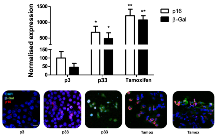
Figure 1. Expression of cellular senescence markers (p16 and β-gal) on P3, P33 and tamoxifen-treated BV2 cells.
Elevated levels of pro-inflammatory markers such as iNOS were also observed in P33 and tamoxifen-treated BV2 cells, showing the effect due to age. Levels of other markers (Arg1, PDL1 or CD80) did not change as much, although the treatment with tumour conditioned mediums from Gl261 and EO771 cells did alter the expression levels of several molecules. These latest results suggest tumour microenvironment may modulate microglia phenotype.
In vivo studies performed displayed that glioblastoma (GL261) and breast cancer (EO771) brain metastasis were significantly decreased in 18 and 24-months-old mice, compared with young 12-weeks mice (Figure 2A). Importantly, iNOS expression in TAMs was significantly increased in aged(24-month-old) animals, whilst the key immunosuppressive marker Arginase- 1 was significantly reduced (Figure 2B). A tumour-suppressing response of microglia in aged animals, showing a closest pro-inflammatory phenotype, can then be suggested.
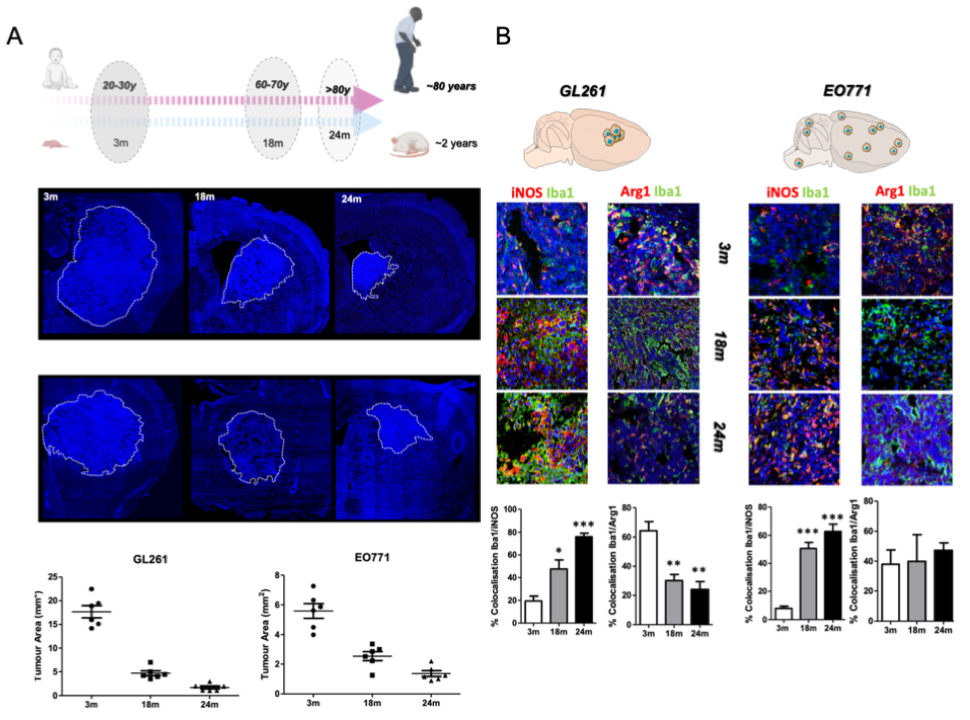
Figure 2. In vivo study of tumour area of glioblastoma and breast cancer brain metastasis at different ages (A). In vivo study of pro- and anti-inflammatory microglial marker levels in glioblastoma and breast cancer brain metastasis at different ages (B).
Conclusion
Cellular senescence characterises aged microglia cells, being one of the main features of this biological process. Aged microglia showed a pro-inflammatory state both, in vitro and in vivo, along with a reduction of anti-inflammatory markers. Altogether, in vivo studies confirm that this senescent-related activation reduced the immunosuppressive tumour microenvironment, supporting brain tumours growth arrest.
Further experiments should focus on understanding better the communication between cancer cells and TAMs on the tumour microenvironment. Promising therapeutic targets for the treatment of brain tumours may emerge from these studies.
References
López-Otín, C., Blasco, M. A., Partridge, L., Serrano, M., & Kroemer, G. (2013). The hallmarks of aging. Cell, 153(6), 1194–1217. https://doi.org/10.1016/j.cell.2013.05.039
Forrester, S. J., Kikuchi, D. S., Hernandes, M. S., Xu, Q., & Griendling, K. K. (2018). Reactive OxygenSpecies in Metabolic and Inflammatory Signaling. Circulation research, 122(6), 877– 902. https://doi.org/10.1161/CIRCRESAHA.117.311401
Di Micco, R., Krizhanovsky, V., Baker, D., & d’Adda di Fagagna, F. (2021). Cellular senescence in ageing: from mechanisms to therapeutic opportunities. Nature reviews. Molecular cell biology, 22(2), 75–95. https://doi.org/10.1038/s41580-020-00314-w
Sousa, A. M. M., Meyer, K. A., Santpere, G., Gulden, F. O., & Sestan, N. (2017). Evolution of the Human Nervous System Function, Structure, and Development. Cell, 170(2), 226–247. https://doi.org/10.1016/j.cell.2017.06.036
Colonna, M., & Butovsky, O. (2017). Microglia Function in the Central Nervous System During Health and Neurodegeneration. Annual review of immunology, 35, 441–468. https://doi.org/10.1146/annurev-immunol-051116-052358
Boza-Serrano, A., Ruiz, R., Sanchez-Varo, R., García-Revilla, J., Yang, Y., Jimenez-Ferrer, I., Paulus,A., Wennström, M., Vilalta, A., Allendorf, D., Davila, J. C., Stegmayr, J., Jiménez, S., Roca-Ceballos,M. A., Navarro-Garrido, V., Swanberg, M., Hsieh, C. L., Real, L. M., Englund,
E., Linse, S., … Deierborg, T. (2019). Galectin-3, a novel endogenous TREM2 ligand, detrimentally regulates inflammatory response in Alzheimer’s disease. Acta neuropathologica, 138(2), 251–273. https://doi.org/10.1007/s00401-019-02013-z
Andreou, K. E., Soto, M. S., Allen, D., Economopoulos, V., de Bernardi, A., Larkin, J. R., & Sibson, N. R. (2017). fAlanmti-minatory Microglia/Macrophages As a Potential Therapeutic
Target in Brain Metastasis. Frontiers in oncology, 7, 251.
Best Practices to Ensure the Wellbeing of Autistic Air Travelers
Namrata (Sethi), A.A.
Presenter
namrata.sethi@haaga-helia.fi
Haaga-Helia University of Applied Sciences, Finland
Katarzyna (Zoltek), B.B.
Haaga-Helia University of Applied Sciences, Finland
Key words: air traveling, autism, wellbeing

Background of the Study/Literature Review
Autism is defined in the psychiatric literature as a neurodevelopmental disorder characterized by a failure on the part of the affected person to communicate and interact socially with others. Autistic persons commonly demonstrate restricted, repetitive, and stereotyped patterns of behaviour. Autism is found in individuals across the world and has no specific propensity for any race, culture, or economic status. It is four times more common in males than in females and is usually diagnosed in childhood when parents and teachers observe that the affected individual fails to make eye contact and interact normally with others. (Autism: A Spectrum Disorder, Joseph S. Alpert, MD, November 09, 2020)
According to the research conducted by Autism Europe, autism spectrum disorder affects around 1 in 100 people in Europe and this rate increases day by day. (Autism Europe, Prevalence rate of autism)
Autism spectrum disorder (ASD) is a broad category with three different levels to specify the degree of support a person needs. ASD is now the umbrella term for the group of complex neurodevelopmental disorders that make up autism. It is a condition that affects communication and behavior.
The autism spectrum refers to the variety of potential differences, skills, and levels of ability that are present in autistic people. Autistic people can experience the following challenges: having trouble communicating and interacting with others, exhibiting repetitive behaviors, having difficulty functioning in several areas of their life (Medical news Today) Autism Spectrum Disorder (ASD) is approached as an invisible disability or hidden disability.
The challenges that ASD travellers face may not seem as obvious to other passengers and staff as those faced by people with a visible disability. However, for an autistic child or adult, air travel can cause various types of stress, including disruption to routines, navigating unfamiliar environments and an overload of sensory stimulation, from loud noises and popping ears to strange smells.
As the aviation industry continues to grow and air travel becomes increasingly popular, it is essential to address the unique challenges and triggers that individuals with autism may experience during their travels. This paper aims to explore the definition, condition, history, types, and characteristics of autism, as well as the impact of autism as a condition on travel behaviour.
When an autistic individual commences their air travel, the disruption to their established routines can be particularly distressing. Autistic individuals often thrive on predictability and sameness, finding comfort and security in the familiarity of their daily routines. The unfamiliar nature of air travel and the processes can provoke anxiety and uncertainty, as autistic individuals strive to adapt to the new and unfamiliar circumstances encountered throughout their journey.
Moreover, the unfamiliarity of airports and aircraft can further compound the stress experienced by autistic travellers. Airports are bustling, dynamic environments characterized by continuous movement, crowds, and a multitude of sensory stimuli. The unfamiliar and chaotic surroundings can make it challenging for autistic individuals to maintain their focus, process information, and navigate through the airport with ease.
Sensory stimulation also plays a significant role in the difficulties faced by autistic individuals during air travel. The sensory-rich environment of airports and aircraft introduces an array of stimuli that can be overpowering for individuals with autism.
Although the challenges faced by autistic travellers during air travel may not always be immediately apparent, they are no less significant. Disruptions to routines, navigating unfamiliar environments, and sensory overload can all contribute to the stress experienced by autistic individuals throughout their journey. By recognizing and addressing these challenges, the aviation industry can foster an environment of inclusivity and support for individuals with autism, ensuring that air travel becomes a more accessible and comfortable experience for them and their families.
Aviation Industry
The aviation industry is one of the fastest-growing industry, it is one of the most popular modes of transportation, providing a unique environment that poses specific challenges for autistic individuals.
However, within this industry lies a distinct environment that presents unique challenges for individuals on the autism spectrum. The sensory-rich and unpredictable nature of air travel has the potential to overwhelm and distress individuals with autism that is why it crucial to develop strategies and best practices to ensure their wellbeing and comfort throughout their journey.
As air travel continues to flourish, it is essential to recognize and address the specific challenges faced by autistic individuals to make travel more inclusive. The unpredictability of the travel experience, combined with the sensory overload, has the potential to provoke heightened anxiety and discomfort.
To mitigate these challenges, it is crucial to implement strategies and best practices that prioritize the well-being and comfort of autistic individuals. By developing a comprehensive understanding of their needs, the aviation industry can create an inclusive and supportive environment. This includes educating airline staff and airport personnel about autism and providing them with the tools and knowledge necessary to effectively assist and accommodate autistic passengers.
Aim of the Study
The aim of this study is to identify and present best practices that airlines and airports can implement to enhance the travel experience of autistic individuals. This paper also emphasizes the importance of understanding the customer journey of autistic travellers and making appropriate adjustments to meet their specific needs.
Methodology
This study employs a comprehensive approach that includes a review of existing literature on autism, travel behaviour, and related challenges. Data collection involves a thorough analysis of academic papers, industry reports, and guidelines concerning autism and travel.
Result/Findings and Argumentation
The findings of this study reveal that autistic individuals face unique challenges in air travel due to their sensory sensitivities, communication difficulties, and reliance on routine and predictability. These challenges can lead to increased anxiety, stress, and potential meltdowns during the travel process. However, by adopting best practices such as providing clear and visual communication, offering pre-flight and in-flight support, training the aviation staff in autism awareness, and creating sensory-friendly environments, airlines and airports can significantly improve the travel experience for autistic individuals. The study identified five best practices which help smooth travel for Autistic air-travellers.
The five best practices below which help ensuring a smoother travel experience for autistic individuals. These practices have been identified as effective tools and strategies that contribute to their comfort and overall well-being during air travel:
- Sunflower lanyard:
The Hidden Disability Lanyard, also known as the sunflower lanyard, has become a widely recognized symbol of hidden disabilities, including autism. By wearing the lanyard, individuals with autism can discreetly indicate to airport staff that they may require additional assistance or understanding. However, it is important to note that while the lanyard serves as a supportive tool, it is essential to confirm and communicate specific assistance requirements with the airport staff well in advance of the journey.
- Autism Visual Guide: An autism visual guide is a valuable resource that provides individuals with autism and their families a comprehensive understanding of the airport environment, procedures, and potential sensory challenges they may encounter during their travel. These guides typically include visual representations, step-by-step instructions, and social stories that help individuals prepare for and navigate the airport experience. By familiarizing themselves with the layout, processes, and potential triggers, autistic individuals can feel more confident and better equipped to manage their journey.
- Separate designated waiting areas: Designated waiting areas specifically designed for autistic individuals before boarding and near immigration checkpoints offer a controlled and quieter environment. These areas provide a space where autistic passengers can wait comfortably, away from the crowds and noise that can be overwhelming. By offering a separate designated space, airports acknowledge the need for a calmer atmosphere that helps reduce anxiety and sensory overload before important stages of the travel process.
- Training for airport staff: Providing training to airport staff on how to effectively assist and interact with autistic passengers is crucial in creating a supportive and inclusive travel experience. This training equips staff members with the knowledge and awareness necessary to understand the unique needs and potential challenges faced by individuals on the autism spectrum. By being educated on autism, airport staff can offer appropriate assistance, demonstrate empathy, and respond to the specific requirements of autistic travellers with greater understanding and sensitivity.
- The Hidden Disability Program is another crucial best practice that aids in smoothening travel for autistic individuals. This program aims to provide discreet support and assistance to passengers with hidden disabilities, including autism. Through the program, airports and airlines collaborate to create a system that recognizes and addresses the specific needs of individuals with hidden disabilities.
These best practices demonstrate the importance of proactive measures in the aviation industry to ensure the well-being and comfort of autistic individuals during their travel. By implementing the use of sunflower lanyards, providing autism visual guides, establishing separate waiting areas, and offering training programs for airport staff, airports can create an environment that is more inclusive, accommodating, and supportive. These practices contribute to a smoother travel experience for autistic individuals and their families, promoting accessibility and equality within the aviation industry.
Conclusion, Managerial Implications, and Limitations
This paper emphasizes the significance of implementing best practices to ensure the wellbeing and comfort of autistic air travellers. The recommendations put forth in this research can serve as a valuable resource for airline and airport management, enabling them to create inclusive environments and enhance the overall travel experience for individuals with autism. It is also essential to acknowledge the limitations of this study, such as the need for further research to assess the effectiveness of these practices and the necessity of considering individual variations within the autism spectrum.
References
Alpert, J. S., MD (2020, November 9). Autism: A Spectrum Disorder. Https://www.Amjmed.com/. Retrieved May 20, 2023, from https://www.amjmed.com/article/S0002-9343(20)30962-1/fulltext
(n.d.). Prevalence rate of autism. Https://www.Autismeurope.org. Retrieved May 20, 2023, from https://www.autismeurope.org/about-autism/prevalence-rate-of-autism/
Klein, A., PsyD (2023, April 23). What are the types of autism? Https://www.Medicalnewstoday.com. Retrieved May 20, 2023, from https://www.medicalnewstoday.com/articles/types-of-autism
Effect of apocarotenoids on β-Amyloid model in Caenorhabditis elegans as a proxy for Alzheimer disease
Zamorano-Aguilar, P.1
Corresponding author – presenter
pedro.zamorano.a@gmail.com
Mapelli-Brahm, P.1
Olmedo, M.2
Meléndez-Martínez, A.J.1
1Food Colour and Quality Laboratory, Facultad de Farmacia, Universidad de Sevilla, 41012, Seville, Spain.
2Departmento de Genética, Facultad de Biología, Universidad de Sevilla, Avenida Reina Mercedes s/n, 41012, Seville, Spain.
Keywords: apocarotenoids, Caenorhabditis elegans, nutraceuticals, functional foods, silver economy.
Background of the study/literature review
In recent decades, there has been a growing interest related to bioactive compounds present in food and their impact on human health. Standing out, carotenoids and their derivatives, apocarotenoids,which are important compounds in ecology, agriculture, aquaculture, nutrition, and health (Meléndez-Martínez et al., 2022). In fact, various studies have reported that some apocarotenoids such as α-ionone, β-ionone, crocin, abscisic acid, among others, exhibit antioxidant, anti-inflammatory, regulatory and signalling properties, to name a few, which contribute to reducing the risk of suffering diseases such as different types of cancer (human gastric adenocarcinoma, breast, prostate andcolon cancer, leukemia), type II diabetes, cardiovascular diseases. And even neurological or metabolic disorders, among others (Ahrazem et al.,2022; Shen et al., 2018; Harrison and Quadro, 2018; Zocchi et al., 2017). For this reason, carotenoids are established as important compounds with great projection to promote health (Meléndez-Martínez, 2019). In fact, the consumption of foods rich in carotenoids and apocarotenoids has been directly related to an improvement at the cognitive level and functioning of the nervous system (Harrison and Quadro, 2018). Thus, some studies have determined that these compounds may have neuroprotective effects, and also improve cognitive function, including memory and mental performance (D’Onofrio etal., 2021; Liao et al., 2023). This, in the context of the aging of the human population and the increase in life expectancy, would allow to increase cognitive performance in advanced age, improving quality of life and promoting healthy aging. The promotion of a healthy diet and a balanced intake of supplements and bioactive compounds such as apocarotenoids, could prevent chronic diseases, and therefore would bring about a significant decrease in medical expenses, both for citizens and for healthcare systems. Since, in the long term, medical visits, hospitalizations, consumption of medicines, procedures, expensive and prolonged treatments would be reduced. As well as the medical personnel necessary for such care would be reduced. In addition, the frequency of medical emergencies due to complications (Heo, 2023; Shanahan and de Lorimier, 2016). On the other hand, the fact that people live longer, and healthier lives has the potential to be positive for society and the economy, since greater labour participation and productivity could be observed (Scott, 2021).
It is worth noting that, besides the positive impact on health, the consumption of functional foods, including those containing apocarotenoids, can have favorable implications at the economic, social, and community development levels, particularly within the emerging segment known as the silver economy (Podgórniak-Krzykacz et al., 2020; Scott, 2021).
Consequently, with the increasing demand for functional foods among the elderly population, numerous opportunities would arise for the agri-food sector. This can be achieved through the development of products enriched with carotenoids, apocarotenoids, and other bioactive compounds, thereby fostering innovation and the growth of functional food offerings. Additionally, it would lead to the creation of new markets and a greater number of jobs in the retail sector and associated services involved in the marketing and distribution of these products.
Among the diseases that significantly impact the quality of life and well-being of the elderly and their families, Alzheimer’s disease stands out as the most prevalent progressive neurodegenerative disorder worldwide. It is characterized by generating behavioral and neuropsychiatric changes, progressive memory loss, mental decline, and cognitive decline (Förstl and Kurz, 1999; Shen et al., 2018). According to the World Alzheimer Report, Alzheimer’s disease is the seventh leading cause of worldwide mortality and affects 55 million people globally (Alzheimer´s Disease International 2021). It is the main cause of dementia in older age, with a predicted prevalence of more than 66 million people over the next 10 years (Markaki and Tavernarakis, 2020). The brains of people with Alzheimer´s disease present neuronal loss in the neocortex accompanied by the accumulation of senile plaques primarily composed of toxic forms of the β-amyloid (Aβ) peptide, particularly Aβ1–42. In fact, the cascade mechanism suggests that abnormal accumulation of the Aβ peptide attend as the initial trigger for Alzheimer´s pathology (Goate et al., 1991; Levy-Lahad et al., 1995; Sherrington et al., 1995; Van Pelt and Truttmann, 2020).
In this regard, Caenorhabditis elegans has emerged as a prominent model for biological and biomedical research, contributing to the understanding of various complex human diseases, includingAlzheimer’s, Parkinson’s, diabetes, cardiovascular diseases, hypertension, and cancer (Baumeister and Ge, 2002; Lublin and Link, 2013; Markaki and Tavernarakis, 2020; Shen et al., 2018). It is worth noting that the C. elegans model has played a crucial role in pivotal discoveries such as RNA interference and apoptosis mechanisms, which have led to Nobel Prizes. Moreover, it is widely employed in biomedicine due to the high degree of conservation of disease pathways between this model and mammalians, including humans (Kaletta and Hengartner, 2006; Liao, 2018). It is estimated that 60-80% of the nematode genes have a human counterpart (Liao, 2018; Markaki and Tavernarakis, 2010). Furthermore, it has been reported that over 83% of the worm proteome presents human homologues, and studies at the genomic level confirm that C. elegans has counterparts for~65% human disease genes (Lai et al., 2000). According to Kamath et al., (2003), at least 1170 genes are essential in C. elegans, as revealed from genome-wide functional analysis (using RNAi) studies. Qin et al., (2018) carried out functional analysis of 143 essential genes, of which 108 were human orthologs. Of these, 89.8% (97 genes) were related to 1218 different diseases (Zhang et al., 2020).
On the other hand, under laboratory conditions, the worm provides numerous advantages. C. elegans can be easily and cost-effectively grown and maintained on agar plates inoculated with the standardnon-pathogenic Escherichia coli strain OP50. It presents a short reproductive cycle (3 – 3.5 days at 20 ºC), short lifespan (2-3 weeks for wild type at 20 ºC), and different organs and tissues (Markaki and Tavernarakis, 2010; Meneely et al., 2019). A single self-fertilizing hermaphrodite can give rise to 300progenies. Therefore, in one week (two generations), it can produce approximately 90,000 – 100,000 offspring (Johnson, 2003; Meneely et al., 2019). These features offer multiple benefits, one of them being that they allow the large-scale production of organisms. Also, it has a complete digestive tract and a nervous system that is widely studied and known. Its connectome or neural map is also available (Varshney et al. 2011). In relation to its immune system, presents relevant similarities with that of mammals (Anastassopoulou et al., 2011; Arvanitis et al., 2013). The genome of C. elegans comprises about 100 million base pairs (Liao, 2018) and was the first among multicellular organism genomes that was completely sequenced and well annotated (The C. elegans Sequencing Consortium, 1998). Besides, it has a transparent body that allows researchers to observe their internal structures and processes indifferent stages of life without bleaching methods. Another key advantage over other models comes from its regulatory status. Since, for European regulations (The European Parliament and the Council of the European Union, 2010), the United States Government (U.S. Government Office of the LawRevision Counsel, 2018), and the Senate of Canada (Senate of Canada, 2015), C. elegans is not legally defined as an animal, which excludes it from ethical prohibitions and limitations.
In relation to Alzheimer’s disease, various transgenic strains that express human pathologic proteins have been developed to imitate the role of Aβ in the development of Alzheimer’s disease (Shen et al., 2018). In this sense, transgenic strains, such as the GMC101 strain, when subjected to thermal stress, have the capacity to express and aggregate β-amyloid (Aβ) Aβ1-42 peptide in body-wall muscle cells (McColl et al., 2012). What to a greater accumulation and concentration results in a progressive or total paralysis.
Aim
Considering the Caenorhabditis elegans model’s ability to investigate Aβ aggregation induced by temperature using transgenic strains, such as strain GMC101, which expresses Aβ in muscle cells and leads to paralysis or loss of movement, this study aimed to evaluate the protective effect of the apocarotenoids α-ionone and β-cyclocitral against Aβ-induced paralysis, serving as a proxy for Alzheimer’s disease in humans. Simultaneously, it seeks to contribute to the interdisciplinary field of nutritional cognitive neuroscience research, emphasizing the potential of dietary components to enhance attention, memory, functional capacity, and overall brain health. This would make it possible to prevent and/or delay age-related neurocognitive deterioration and impairments.
Methodology
In this study, C. elegans GMC101 was fed with E. coli OP50 as a control group, while the experimental group received a mixture of E. coli OP50 supplemented with varying concentrations of α-ionone and β-cyclocitral (ranging from 10 to 500 μg/mL). Embryos were hatched in the absence of food to synchronize to the L1 stage and subsequently grown on plates with food supplemented with different carotenoids, at a temperature of 16.5°C for 72 hours until they reached the young adult stage. Then, they were transferred to 25°C to induce β- amyloid (Aβ) aggregation. After 24 hours, the fraction of paralyzed animals was quantified in three plates per experiment. The effects of α-ionone and β-cyclocitral on the cultured worms were analyzed using Dunnett’s test and one-way ANOVA with Prism version 8.0.1 for Windows (GraphPad Software, CA, USA). A significance level of p<0.05 was considered statistically significant.
Results
Both tested compounds demonstrated a significant reduction of the Ab aggregation-induced paralysis, with β-cyclocitral showing the highest impact on C. elegans mobility (Table 1). Its effect displayed a clear dose-response relationship, with the percentage of paralysis gradually decreasing within creasing concentrations of the compounds. For β-cyclocitral, the lowest concentration tested (10 µg/mL) already showed a modest effect on paralysis, with 71% of the worms paralyzed, compared to 75% in the control. Increased concentrations provoked an increasing effect, with the highest concentration (500 µg/mL) resulting in only 46% of paralyzed worms (Fig. 1a). The statistical analysis showed significant differences with the control for the concentrations higher than 100 µg/mL (Fig. 1c; Table 1).
In relation to α-ionone supplementation, the results showed that the greatest anti paralysis effect was obtained at doses of 100 µg/mL and 250 µg/mL. Since, 67% and 57% of the worms were paralyzed (Fig. 1b). On the other hand, compared to 76% of the control, the effect of the other concentrations tested was not significant, since the percentages of paralyzed worms ranged between 72 – 73% (Fig. 1d; Table 1).
Conclusions
Our findings suggest that α-ionone and β-cyclocitral effectively decrease Aβ-induced paralysis in a C. elegans model. These findings provide valuable insights for future investigations and strategies aimed at reducing the aggregation of β-amyloid peptide, potentially benefiting human beings by preventing age-related neurocognitive disorders.
Therefore, this study emphasizes the significance of nutrition and diet, particularly the role of apocarotenoids, in promoting human health. It highlights the interconnectedness between the concepts of functional foods, bioactive compounds, increased life expectancy, and the silver economy, with the ultimate goal of fostering a healthy and sustainable society.
Extra – Figures and Tables
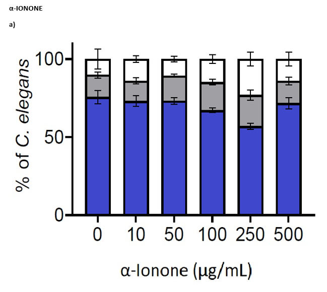
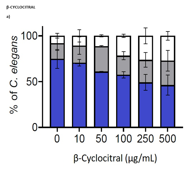
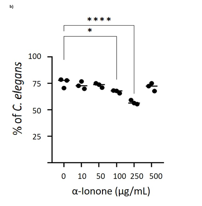
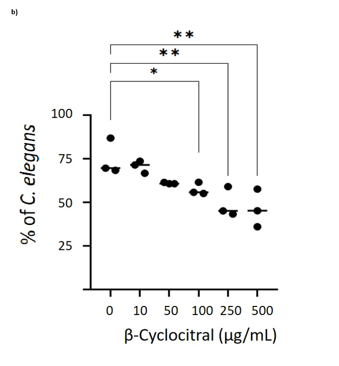
Fig 1. Effect of the E. coli OP50 supplemented with different concentrations of apocarotenoids (0 (control), 10, 50, 100, 250, 500 µg/mL) in adult GMC101 C. elegans strain. Percentage of movement, semi-paralysis and paralysis with (a) α-Ionone, and (b) β-cyclocitral. Worms paralyzed average with (c) α-ionone, and (d) β-cyclocitral. The number of paralyzed worms was scored 24 h after heat shock, at adult stage. Data are show as percentage ± SD of at least 100 nematodes in 3 independent biological replicates (in triplicate). Statistical significance was determined by Dunnett´s test with results compared with the control group. p<0.05 was taken as statistically significant.
Table 1. Anti-paralysis effect of apocarotenoids in GCM C. elegans strain.
| Apocarotenoid | Concentration (µg/mL) | % paralysis | P value | Statistical difference |
| α-ionone | 10 | 73.03 | 0.7628 | No |
| 50 | 73.18 | 0.8002 | No | |
| 100 | 67.14 | 0.0210 | Yes (*) | |
| 250 | 56.92 | <0.0001 | Yes (****) | |
| 500 | 71.61 | 0.4072 | No | |
| Control | 75.56 | |||
| β-cyclocitral | 10 | 70.53 | 0.9194 | No |
| 50 | 60.87 | 0.1334 | No | |
| 100 | 57.39 | 0.0495 | Yes (*) | |
| 250 | 49.08 | 0.0043 | Yes (**) | |
| 500 | 46.20 | 0.0019 | Yes (**) | |
| Control | 74.86 |
References
Ahrazem, Oussama, Changfu Zhu, Xin Huang, Ángela Rubio-Moraga, Teresa Capell, Paul Christou, and Lourdes Gómez-Gómez. 2022. “Metabolic Engineering of Crocin Biosynthesis in Nicotiana Species.” Frontiers in Plant Science 13 (March): 861140. https://doi.org/10.3389/fpls.2022.861140.
Alzheimer’s Disease International. 2021. World Alzheimer Report 2021: Journey throughthe diagnosis of dementia. https://www.alzint.org/resource/world-alzheimer-report-
Anastassopoulou, C. G., B. B. Fuchs, and E. Mylonakis. 2011. Caenorhabditis elegans- based Model Systems for Antifungal Drug Discovery. Current Pharmaceutical Design, 17 (13):1225–1233.
Arvanitis, M., D. D. Li, K. Lee, and E. Mylonakis. 2013. Apoptosis in c.elegans: Lessons for cancer and immunity. Frontiers in Cellular and Infection Microbiology, 3:67. https://doi.org/10.3389/fcimb.2013.00067
Baumeister, Ralf, and Liming Ge. 2002. “Baumeister, R. & Ge, L. The Worm in Us – Caenorhabditis Elegans as a Model of Human Disease. Trends Biotechnol. 20, 147-148.” Trends in Biotechnology 20 (May): 147–48. https://doi.org/10.1016/S0167-7799(01)01925-4.
D’Onofrio, Grazia, Seyed Nabavi, Daniele Sancarlo, Antonio Greco, and Stefano Pieretti. 2021. “Crocus Sativus L. (Saffron) in Alzheimer’s Disease Treatment: Bioactive Effects on Cognitive Impairment.” Current Neuropharmacology 19 (January). https://doi.org/10.2174/1570159X19666210113144703.
Förstl, H., and Kurz, A. 1999. Clinical features of Alzheimer´s disease. European Archives of Psychiatry and Clinical Neuroscience, 249(6), 288–290.
Goate, A., Chartier-Harlin, M.-C., Mullan, M., Brown, J., Crawford, F., Fidani, L., Giuffra, L., Haynes, A., Irving, N., James, L., et al. 1991. Segregation of a missense mutation in the amyloid precursor protein gene with familial Alzheimer’s disease. Nature, 349(6311), 704–706. https://doi.org/10.1038/349704a0
Harrison, Earl, and Loredana Quadro. 2018. “Apocarotenoids: Emerging Roles in Mammals.” Annual Review of Nutrition 38 (August). https://doi.org/10.1146/annurev-nutr- 082117-051841.
Heo, Seok-Hyun. 2023. “A Study Is Needed to Investigate the Influence of Health FunctionalFoods on Reducing National Medical Expenses.” Food Suppl Biomater Health 3
(1). <ahref=”https://doi.org/10.52361/fsbh.2023.3.e2″>https://doi.org/10.52361/fsbh.2023.3.e2.
Johnson, T. E. 2003. Advantages and disadvantages of Caenorhabditis elegans for aging research. In Experimental Gerontology, 38 (11-12):1329–1332. Elsevier Inc. https://doi.org/10.1016/j.exger.2003.10.020
Kaletta, Titus, and Michael Hengartner. 2006. “Kaletta, T. & Hengartner, M.O. Finding Function in Novel Targets: C. Elegans as a Model Organism. Nat. Rev. Drug Discov. 5, 387- 398.” Nature Reviews. Drug Discovery 5 (June): 387–98. https://doi.org/10.1038/nrd2031.
Kamath, R. S., A. G. Fraser, Y. Dong, G. Poulin, R. Durbin, M. Gotta, A. Kanapink, N. le Bot, S. Moreno, M. Sohrmann, et al. 2003. Systematic functional analysis of the Caenorhabditis elegans genome using RNAi. Nature, 421:231–237. www.nature.com/nature
Lai, C. H., C. Y. Chou, L. Y. Ch’ang, C. S. Liu, and W. C- Lin. 2000. Identification of Novel Human Genes Evolutionarily Conserved in Caenorhabditis elegans by Comparative Proteomics. Genome Research, 10 (5):703–713. www.genome.org
Levy-Lahad, E., Wasco, W., Poorkaj, P., Romano, D. M., Oshima, J., Pettingell, W. H., Yu, C., Jondro, P. D., Schmidt, S. D., Wang, K., et al. 1995. Candidate Gene for the Chromosome 1 Familial Alzheimer’s Disease Locus. Science, 269(5226), 973–977. http://www.jstor.org/stable/2887712
Liao, Ping, Qing-Yun Wu, Sen Li, Kai-Bin Hu, Hui-Lin Liu, Hai-Yan Wang, Zai-Yun Long, Xiu-Min Lu, and Yong-Tang Wang. 2023. “The Ameliorative Effects and Mechanisms of Abscisic Acid on Learning and Memory.” Neuropharmacology 224: 109365. https://doi.org/https://doi.org/10.1016/j.neuropharm.2022.109365.
Liao, Vivian Hsiu-Chuan. 2018. “Use of Caenorhabditis Elegans To Study the Potential Bioactivity of Natural Compounds.” Journal of Agricultural and Food Chemistry 66 (8): 1737–42. https://doi.org/10.1021/acs.jafc.7b05700.
Lublin, A L, and C D Link. 2013. “Alzheimer’s Disease Drug Discovery: In Vivo ScreeningUsing Caenorhabditis Elegans as a Model for β-Amyloid Peptide-Induced Toxicity.” Drug Discovery Today: Technologies 10 (1): e115–19. https://doi.org/https://doi.org/10.1016/j.ddtec.2012.02.002.
Luyten, Walter, Peter Antal, Bart P Braeckman, Jake Bundy, Francesca Cirulli, ChristopherFang-Yen, Georg Fuellen, et al. 2016. “Ageing with Elegans: A Research Proposal to Map Healthspan Pathways.” Biogerontology 17 (4): 771–82. https://doi.org/10.1007/s10522-016-9644-x.
Markaki, M., and N. Tavernarakis. 2010. Modeling human diseases in Caenorhabditis elegans.In Biotechnology Journal, 5(12): 1261–1276. https://doi.org/10.1002/biot.201000183
Markaki, M, and N. Tavernarakis. 2020. “Caenorhabditis Elegans as a Model System for Human Diseases.” Current Opinion in Biotechnology 63 (January): 118–25. https://doi.org/10.1016/j.copbio.2019.12.011.
Mccoll, G., Roberts, B. R., Pukala, T. L., Kenche, V. B., Roberts, C. M., Link, C. D., Ryan, T. M., Masters, C. L., Barnham, K. J., Bush, A. I., et al. 2012. Utility of an improved model of amyloid-beta (Aβ 1-42 ) toxicity in Caenorhabditis elegans for drug screening for Alzheimer’s disease. Molecular Neurodegeneration, 7, 57. http://www.molecularneurodegeneration.com/content/7/1/57
Meléndez-Martínez, Antonio J. 2019. “An Overview of Carotenoids, Apocarotenoids, and Vitamin A in Agro-Food, Nutrition, Health, and Disease.” Molecular Nutrition & Food Research 63 (15): 1801045. https://doi.org/https://doi.org/10.1002/mnfr.201801045.
Meléndez-Martínez, Antonio J, Anamarija I Mandić, Filippos Bantis, Volker Böhm, Grethe Iren A Borge, Mladen Brnčić, Anette Bysted, et al. 2022. “A Comprehensive Review on Carotenoids in Foods and Feeds: Status Quo, Applications, Patents, and Research Needs.” Critical Reviews in Food Science and Nutrition 62 (8): 1999–2049. https://doi.org/10.1080/10408398.2020.1867959.
Meneely, P. M., C. L. Dahlberg, and J. K. Rose. 2019. Working with Worms: Caenorhabditis elegans as a Model Organism. Current Protocols Essential Laboratory Techniques, 19 (1), e35. https://doi.org/https://doi.org/10.1002/cpet.35
Podgórniak-Krzykacz, Aldona, Justyna Przywojska, and Izabela Warwas. 2020. “Silver Economy as a Response to Demographic Challenges in Polish Regions: Realistic Strategy or Utopia?” Innovation: The European Journal of Social Science Research, March, 1–28. https://doi.org/10.1080/13511610.2020.1736011.
Qin, Z., R. Johnsen, S. Yu, J. S. C. Chu, D. L. Baillie, and N. Chen. 2018. Genomic identification and functional characterization of essential genes in Caenorhabditis elegans. G3: Genes, Genomes, Genetics, 8 (3):981–997. https://doi.org/10.1534/g3.117.300338
Scott, Andrew. 2021. “The Longevity Society.” The Lancet Healthy Longevity 2 (December): e820–27. https://doi.org/10.1016/S2666-7568(21)00247-6.
Senate of Canada. 2015. BILL S-214: An Act to amend the Food and Drugs Act (cruelty- free cosmetics). Https://Www.Parl.ca/LegisInfo/En/Bill/42-1/s-214.
Shanahan, Christopher J, and Robert de Lorimier. 2016. “From Science to Finance—A Tool for Deriving Economic Implications from the Results of Dietary Supplement Clinical Studies.” Journal of Dietary Supplements 13 (1): 16–34. https://doi.org/10.3109/19390211.2014.952866.
Shen, Peiyi, Yiren Yue, Jolene Zheng, and Yeonhwa Park. 2018. “Caenorhabditis Elegans: AConvenient In Vivo Model for Assessing the Impact of Food Bioactive Compounds on Obesity, Aging,and Alzheimer’s Disease.” Annual Review of Food Science and Technology 9 (March). https://doi.org/10.1146/annurev-food-030117-012709.
Sherrington, R., Rogaev, E. I., Liang, Y., Rogaeva, E. A., Levesque, G., Ikeda, M., Chi, H., Lin, C., Li, G., Holman, K., et al. 1995. Cloning of a gene bearing missense mutations in early-onset familial Alzheimer’s disease. Nature, 375(6534), 754–760. https://doi.org/10.1038/375754a0
The C. elegans Sequencing Consortium. 1998. Genome Sequence of the Nematode C. elegans: A Platform for Investigating Biology. https://doi.org/10.1126/science.282.5396.2012
The European Parliament and the Council of the European Union. 2010. DIRECTIVE 2010/63/EU OF THE EUROPEAN PARLIAMENT AND OF THE COUNCIL of 22 September 2010 on the protection of animals used for scientific purposes
U.S. Government Office of the Law Revision Counsel. 2018. 7 USC Ch. 54: TRANSPORTATION, SALE, AND HANDLING OF CERTAIN ANIMALS. Https://Uscode.House.Gov/View.Xhtml?Path=/Prelim@title7/Chapter54&edition=prelim.
van Pelt, K. M., and Truttmann, M. C. 2020. Caenorhabditis elegans as a model system forstudying aging-associated neurodegenerative diseases. Translational Medicine of Aging, 4, 60–72. https://doi.org/10.1016/j.tma.2020.05.001
Varshney, L. R., B. L. Chen, E. Paniagua, D. H. Hall, and D. B. Chklovskii. 2011. Structural properties of the Caenorhabditis elegans neuronal network. PLoS Computational Biology, 7 (2), e1001066. https://doi.org/10.1371/journal.pcbi.1001066
Zhang, S., F. Li, T. Zhou, G. Wang, and Z. Li. 2020. Caenorhabditis elegans as a Useful Model for Studying Aging Mutations. Frontiers in Endocrinology, 11, 554994. https://doi.org/10.3389/fendo.2020.554994
Zocchi, Elena, Raquel Hontecillas, Andrew Leber, Alexandra Einerhand, Adrià Carbó, Santina Bruzzone, Nuria Tubau-Juni, et al. 2017. “Abscisic Acid: A Novel Nutraceutical for Glycemic Control.” Frontiers in Nutrition 4 (June). https://doi.org/10.3389/fnut.2017.00024.
“Exploring the Link Between Blue-Green Spaces and Well-being: A Survey of Shkodra’s Lake, Albania”
Kruja Samel
Corresponding author – presenter
samkru@alum.us.es
University of Seville, Spain
Keywords: Blue space, green space, urban planning, health, well-being

Background
Cities have historically adapted their shape in response to the challenges of public health. Over 60% of the metropolitan areas that will exist by 2050 are expected to be built, implying that enormous additional infrastructure requirements would be required, notably in Asia and Africa (Hartig et al.,2014). At the same time, current cities throughout the world are aging and in desperate need of infrastructural upgrades. On the other hand, urban environments contain facilities that may help in reducing the severe health consequences of city living. Because many chronic diseases are avoidable, there has been a need for reasonable measures taken by population and government to minimize risk or mitigate damages from smoking, physical inactivity, alcohol consuming, and an unhealthy diet (Bauer et al., 2014). Due to the great benefits provided by nature, in both theoretical and practical aspects during the last two decades, there has been an increasing interest in ecological, nature-basedhealth-promoting initiatives (Hartig et al., 2014). The construction, protection, maintenance, and growth of blue and green areas (BGS) are the goals of new strategies.
Interactions with nature have been shown to improve psychological well-being (Kaplan 1995), improve mood (Hartig et al., 2003, Barton & Pretty, 2010, Roe & Aspinall, 2011), increase focus (Hartig et al., 2003; Ottosson & Grahn, 2005), and lower level of stress and anxiety (Hartig et al., 2003; Ottosson & Grahn, 2005). The reasons of poor mental health are numerous and complex (Kinderman et al. 2015), and cultural and socio-economic variations between locations may impact how people respond to natural interactions (Keniger et al., 2013). Nature is likely to have an impact on mental balance by a series of functions (Shanahan et al., 2015b). Green spaces are thought to provide health advantages through encouraging physical exercise, contact with nature, and social interaction, according to a paradigm built based on existing studies (Lachowycz & Jones, 2013).
Even though the relationship among blue and green spaces and human health is complicated in every country, understanding of its dynamics, as well as amounts of information and data on health and well-being, as well as access to green and blue spaces, vary. The goal of this study is to bridge the gap between (1) understanding complex relationship among blue and green spaces, and positive effects on well-being in the Shkodra’ population, and (2) describing its complicated dynamics. In Albania, studies that relate the impact of blue and green spaces on the population’s well-being has been limited and poorly explored.
Methodology
Shkodra is an ancient town of 2500 years old and one of the most important cities of Albania. It is situated in the northwest of Albania, with a surface area of 872,71 km2. According to INSTAT data (2021) on 1st January, the Shkodra’ municipality has a population of 200,007 inhabitants where 48,6% are men and 51,4% are women (INSTAT, 2020). The lake of Shkodra with 369 km2, the largest lake in Albania and Montenegro, is located on the west of the city of Shkodra and serves as a border between the two countries (Sadori et al., 2014), with 149 km of it belongs to Albania. Population data presented in this paper are preliminary data extracted from an online cross-sectional survey on BGS carried out in Shkodra’ city. Respondents have been asked to complete the survey via the platform Google Form, from April to May 2021. During this period, Albania was open to all citizens and visitors, with some restrictions in place, such as masks required outside and inside certain buildings and institutions, no gatherings of more than 50 people, and public movement prohibited from 10:00 p.m. to 5:00 a.m. The questionnaire used in the study was prepared by an interdisciplinary panel formed by urban planner, geographer, psychologist, and environmental scientist. The questionnaire provided 68 questions, designed in 3 main sections: 1) General information, 2) Natural environment information, 3) Self-reported health information. To improve the clarity of the questions before launching the survey, a pilot study was conducted. After the validation and cleaning process, a representative sample (95% level of confidence) of 530 respondents was obtained. This survey targeted people over 16 years old and was disseminated to the public using social media platforms.
Descriptive statistics were used to analyze such as indicators (1) sociodemographic characteristics (age, gender, marital status, level of education and job status, years living in Shkodra; (2) frequency of visits in BGS in the last 4 weeks; (3) time spending during the visit; (4) activities carried out during the visit; (5) type of accompaniment; (6) the reason for not visiting BGS and the quality of BGS. The SPSS software platform was used for the statistical analyses. The frequency, percentage, mean and standard deviation calculations were used to calculate data from the sample. The Chi-square test was used to analyze association of visits frequency in blue-green space with the people’ mood as an indicator of well-being. There exists any statistical significance when p-value was P <0.05.
Results
According to the analysis of the sociodemographic variables, the mean age of the respondents was 30.32 ± 12.971. It can be observed that the population sample was young, with 63.4% between 16 and 31 years old, 23.4% between 32 and 48 years old, 12.6% between 49 and 64 years old, and only 0.6% over 65 years old, including 76.2% of women and 23.8% of men. Regarding employment status,55.8% were working at the time of the survey, 30.2% were students, 1.9% were unemployed, 1.1%were homemakers followed by 0.8% retired, and 0.2% disabled. It is interesting to remark that only 14.7% of the respondents had not visited Shkodra’
Lake during the last four weeks of whom 43.5% for lack of time, 37.1% for living too far from this area, and only 3.8% for describing it as an overpopulated area. In terms of frequency of visits to Shkodra’ Lake, 30.9% had visited once or twice in the last four weeks, 28.1% several times a week, 26.2% had visited only once a week and 14.7% had not made any visits in the last four weeks. Concerning the type of accompaniment during the visits, 37.9% visited Shkodra Lake with friends, 14.2% with wife/husband or boyfriend/girlfriend, 13.4% with children, 8.5% with parents, 5.8% with another adult, and 2.8% alone. In their visits, the respondents spent approximately 60 minutes at the Shkodra’ Lake BGS as follows: 31.1% cycling, 18.3% consuming food and drink, 8.9% walked or played with children, 4.3% engaged in quiet activities (e.g., reading, meditating), 0.8% walked with a dog, 0.6% went swimming and 0.2% used this time for fishing. In terms of quality, 46.6% of the people who had visited during the last 4 weeks rated this area as good quality, 29.8% rated it as acceptable, 14.8% considered Shkodra’ Lake as very good quality and only 7.5% thought that the quality of this area was bad or very bad.
Thus, according to the analysis of the socio-demographic variables, the mean age of the respondents was 30.32 ± 12.971. It can be observed that the population sample was young, with 63.4% be-tween 16 and 31 years old, 23.4% between 32 and 48 years old, 12.6% between 49 and 64 years old, and only 0.6% over 65 years old, including 76.2% of women and 23.8% of men. Regarding employment status, 55.8% were working at the time of the survey, 30.2% were students, 1.9% were unemployed, 1.1% were homemakers followed by 0.8% retired, and 0.2% disabled. It is interesting to remark that only 14.7% of the respondents had not visited Shkodra’ Lake during the last four weeks of whom 43.5% for lack of time, 37.1% for living too far from this area, and only 3.8% for describing it as an overpopulated area. In terms of frequency of visits to Shkodra’ Lake, 30.9% had visited once or twice in the last four weeks, 28.1% several times a week, 26.2% had visited only once a week and 14.7% had not made any visits in the last four weeks. Concerning the type of accompaniment during the visits, 37.9% visited Shkodra Lake with friends, 14.2% with wife/husband or boyfriend/girlfriend, 13.4% with children, 8.5% with parents, 5.8% with another adult, and 2.8% alone. In their visits, the respondents spent approximately 60 minutes at the Shkodra’ Lake BGS as follows: 31.1% cycling, 18.3% consuming food and drink, 8.9% walked or played with children, 4.3% engaged in quiet activities (e.g., reading, meditating), 0.8% walked with a dog, 0.6% went swimming and 0.2% used this time for fishing. In terms of quality, 46.6% of the people who had visited during the last 4 weeks rated this area as good quality, 29.8% rated it as acceptable, 14.8% considered Shkodra’ Lake as very good quality and only 7.5% thought that the quality of this area was bad or very bad.
Conclusions
The current study found a correlation between people’s mood and visits to blue and green spaces(BGS), in a way that people who visited Shkodra’ Lake more frequently demonstrated higher positive feelings. Therefore, the results of this study confirm the results acquired from previous studies claiming the benefits of visits to BGS on mood and well-being of people (White et al., 2013; Su Sugiyama et al., 2008; Pretty et al., 2005).Although there are studies that affirm that visiting green spaces has beneficial impact on physical and mental health (White et al., 2013) and helps us to be more relaxed and less stressed (Kellert, 1995), there is noticeable a lack of studies in the Mediterranean area in general (Braçe et al., 2020) and Albania in particular, that would provide knowledge on the impact of BGS on the population health and well-being.
The results of this study reinforce the importance of BGS as a community resource to promote good health. Therefore, local governments who are often in charge of the design and maintenance of BGS could address community health problems by improving and creating BGS. However, studies have shown that BGSs, in general, are underutilized. Although the health benefits of BGS have been shown, it is likely that such findings have not been adequate to persuade decision-makers to act. It is critical to emphasize that strengthening BGS is probably a practical health promotion effort that local governments can undertake. Although the findings are not conclusive, there is evidence that access to safe, clean, and appealing blue spaces has several health and wellness advantages, due to different processes such as reducing temperatures, increasing physical activity, reducing stress, and promoting quality time with relatives and friends.
References
- Bauer, U. E., Briss, P. A., Goodman, R. A., & Bowman, B. A. (2014). Prevention of chronic disease in the 21st century: elimination of the leading preventable causes of premature death and disability in the USA. The Lancet, 384(9937), 45-52.
- Hartig, T., & Kahn, P. H. (2016). Living in cities, naturally. Science, 352(6288), 938- 940.
- Hartig, T., Evans, G. W., Jamner, L. D., Davis, D. S., & Gärling, T. (2003). Tracking restoration in natural and urban field settings. Journal of environmental psychology, 23(2), 109-123.
- Hartig, T., Mitchell, R., De Vries, S., & Frumkin, H. (2014). Nature and health. Annual review of public health, 35, 207-228.
- Kaplan, S. (1995). The restorative benefits of nature: Toward an integrative framework. Journal of environmental psychology, 15(3), 169-182.
- Keniger, L. E., Gaston, K. J., Irvine, K. N., & Fuller, R. A. (2013). What are the benefits of interacting with nature?International journal of environmental research and public health, 10(3), 913-935.
- Lachowycz, K., & Jones, A. P. (2013). Towards a better understanding of the relationship between greenspace and health: Development of a theoretical framework. Landscape and urban planning, 118, 62-69.
- Shanahan, D. F., Lin, B. B., Bush, R., Gaston, K. J., Dean, J. H., Barber, E., & Fuller, R. A. (2015). Toward improved public health outcomes from urban nature. American journal of public health, 105(3), 470-477.
Folic acid homeostasis and its pathways related to hepatic oxidation inadolescent rats exposed to binge drinking
Gallego-López, M.d.C.
Corresponding author – presenter
mgallego3@us.es
Department of Physiology, Faculty of Pharmacy, University of Seville, Seville, Spain
Ojeda, M.L.; Romero-Herrera, I.
Nogales, F.; Carreras, O.

Background
Chronic ethanol consumption (Chr-EtOH) and alcoholic liver disease (ALD) are intimately related to folic acid (FA) homeostasis, which is well recognized in clinical and experimental research. Therefore, currently, FA is supplied to these patients in order to prevent ALD, as this is the main vitamin deficiency from which they suffer (Medici & Halsted, 2013).
The mechanisms of Chr-EtOH injury are multifactorial, involving several pathways, most of them related to its oxidative metabolism, which takes place mainly in the liver. One of these EtOH metabolizer enzymes is CYP2E1, that directly produces reactive oxygen species (ROS) increasing oxidative stress (Hernández-Rodríguez et al., 2014). Biomolecular oxidative damage is considered one of the main mechanisms related to ALD.
FA, the synthetic form of natural folates, forms part of the water-soluble B vitamins family and is taken up from the diet and bio-transformed mainly in the liver by the enzyme dihydrofolate reductase (DHFR). It functions as a coenzyme in reactions of one-carbon transfer, needed for the biosynthesis of several essential molecules like DNA, RNA and amino acids. Thus, adequate intake of FA is vital for cellular homeostasis, with folates being critical during periods of rapid growth such as adolescence (Navarro-Pérez et al., 2016). In addition, antioxidant properties have been attributed to FA (Hwang et al., 2011; Joshi et al., 2001; Stanhewicz & Kenney, 2017; Woo et al., 2006). However, these studies approach the issue from a one-sided point of view, so there is not a well-elucidated overview of all FA antioxidant mechanisms in the same study.
Lately, adolescents have been practising binge drinking (BD), consisting of the intake of a high amount of alcohol in a short time (de Medeiros et al., 2023; National Institute on Alcohol Abuse and Alcoholism, 2020). This is particularly harmful since adolescents are very vulnerable to the toxic effects of EtOH, and this acute consumption pattern is drastically pro-oxidant. Acute EtOH consumption leads to different biological effects than Chr-EtOH, since in its metabolism, it produces higher amounts of ROS by increasing CYP2E1 activity (Ojeda et al., 2022). Therefore, BD could produce different changes in FA homeostasis, compromising the oxidative balance even more. However, thus far, there are few studies related to acute EtOH consumption and FA homeostasis.
Aim
The aim of this study is to examine, for the first time, FA homeostasis in BD adolescent rats and its antioxidant properties in the liver.
We will obtain a general overview of FA involvement in ROS and reactive nitrogen species over-production, oxidative/nitrosative stress balance and biomolecular damage which could induce apoptosis during BD exposure in adolescence.
Methodology
Four groups of Wistar male adolescent rats were used to develop this experiment: control, BD, control FA-supplemented group and BD FA-supplemented group. Dietary FA content in control groups was 2ppm, and 8 ppm in supplemented groups. EtOH exposed groups received the intermittent alcohol treatment called “binge drinking” by the administration of an intraperitoneal (i.p.) injection of EtOH (3 g/kg) in physiological saline solution (PSS) at 20% (v/v) during 3 consecutive days per week for3 weeks (Callaci et al., 2010). The rest of the groups were administered an equivalent volume of PSS i.p. over the same period. This i.p. forced BD method was chosen in order to avoid digestive interferences with FA oral supplementation and to analyze the direct effect of this drug on the body. Each day during the three weeks of treatment, the food intake (g/day) and the rats’ body weight (g/day) were measured. Animal care procedures and experimental protocols were in accordance with EU regulations (Council Directive 86/609/EEC,24 November 1986) and were approved by the Ethics Committee of the University of Seville (CEEA-US2019-4).
Once the experimental period was over, the animals were humanely sacrificed. Tissue, urine and serum samples were removed for biochemical determinations. Afterwards, FA, glutathione (GSH) and nitric oxide (NO) levels were tested by ELISA; hepatic transaminases were measured with an automated analyser; antioxidant enzymes activity and oxidative stress markers were examined spectrophotometrically; and hepatic proteins expression was carried out by Western blot.
The obtained data were analysed using the analysis of variance (ANOVA) in the program GraphPadInStat3. Subsequently, the Tukey–Kramer test was used to determine the significant differences between the means.
Main findings and argumentation
From a nutritional and morphological point of view, neither i.p. BD exposure nor FA supplementation provoke changes in the studied animals. However, in this study it has been demonstrated that as in Chronic-EtOH patients, BD in adolescence also disrupts FA homeostasis. Renal FA reabsorption and renal FA deposits are increased, indicating that the body is making an effort not to excrete it in urine, increasing its retention. Hepatic deposits are decreased, and heart and serum levels remain unaffected. This depletion of FA in the liver is showing that hepatic damage is taking place since transaminases levels are increased.
Furthermore, oxidative stress after BD exposure is clearly established. BD provokes an important imbalance in the endogenous enzymatic system, the one that is in charge of detoxifying ROS(Hernández-Rodríguez et al., 2014). It increases superoxide dismutase (SOD) and catalase (CAT) activities and decreases glutathione peroxidase (GPx) activity, leading to lipid and protein oxidation and a decrease of the antioxidant GSH levels.
It is also highlighting that, again as in Chronic-EtOH exposure (Vatsalya et al., 2021), BD increases serum homocysteine (Hcy) levels causing hyper-homocysteinemia (HHcy), a dangerous state that is correlated with diverse pathologies, such as vascular dysfunction, neurodegenerative disorders and of course, hepatic injury (Murray et al., 2015). Probably, BD increases Hcy levels by altering the methionine cycle and the transsulfuration pathway, leading to lower GSH synthesis. These effects of EtOH are attributed to the ROS generated, and they are exacerbated by a lack of hepatic FA that serves as a cofactor in these pathways (Dumitrescu, 2018; Vatsalya et al., 2021).
Among other actions, Hcy increases the pro-oxidant nicotinamide adenine dinucleotide phosphate (NADPH) oxidase (NOX) activity (Hung-Chih et al., 2016; Murray et al., 2015). In fact, in BDadolescent rats NOX1 and especially NOX4 expressions are increased, affecting the oxidative state and apoptosis generation since caspase-9 and caspase-3 are also increased in the liver; and it is known that these pro-apoptotic enzymes are increased in cell stress situations (Ting-Jun et al., 2005).
BD also induces nitrosative stress because it increases the expression of uncoupled endothelial nitric oxide synthase (eNOS) (ROS generator), leading to the nitration of tyrosine proteins (protein-SNOs). This situation occurs, in part, since DHFR is decreased in the liver of BD adolescent rats. DHFR is an enzyme that transforms dihydrobiopterin (BH2) into tetrahydrobiopterin (BH4) (Alp & Channon, 2004), the main cofactor for the eNOS dimer/coupled form (NO generator); consequently, in BD exposed rats BH4 is probably decreased, which collaborates in the increase of the uncoupled eNOS form. The enzyme DHFR depends on FA levels, connecting with the lower FA hepatic deposits found in these animals (Gao et al., 2012).
Therefore, in BD adolescent rats FA deposits in the liver are probably being consumed in order to avoid the oxidative, nitrosative and apoptotic BD-EtOH damage in this tissue.
Thus, when a FA supplementation is administered, FA homeostasis is restored, being important the recovery of hepatic levels, which improves liver function by decreasing transaminase levels. FA supplementation also improves the oxidative balance by reducing lipid and protein oxidation by different mechanisms. It decreases SOD activity; an action that could be due to its supposed effects as a scavenger directly decreasing ROS concentration (Joshi et al., 2001); by inducing other antioxidant mechanisms that indirectly decrease ROS generation, such as decreasing NOX activity (Woo et al., 2006); or by decreasing Hcy levels, which stimulates SOD (Hwang et al., 2011). FA supplementation also increases GSH levels, which confirm that hepatic GSH levels during BD in rats are deeply related to FA deposits and it is regenerated mainly by the transsulfuration pathway, which is reestablished after FA supplementation.
As previously anticipated, FA therapy decreases NOX1 and NOX4 expressions and avoids HHcy, contributing to reduce the oxidative and apoptotic state. This is probably due to FA contributing to restoring Hcy remethylation to methionine or to catabolizing it through the transsulfuration pathway, increasing GSH hepatic levels. Then, FA supplementation reduces caspases expression, indicating that apoptosis is decreased when FA hepatic levels are balanced, and that this vitamin plays an important role during BD liver damage.
Regarding the nitrosative state, FA therapy by restoring DHFR expression leads to recoupled eNOSexpression in BD rats. It increases NO production and decreases protein-SNOs in the liver, avoiding nitrosative stress. Different mechanisms are related to FA and eNOS activity in endothelial cells, including greater bioavailability of BH4 through stabilization of BH4 and/or recycling from BH2, mainly by upregulating the activity of DHFR in the biopterin recycling pathway, direct interaction with eNOS, and directly decreasing ROS generation (Stanhewicz & Kenney, 2017).
Conclusion
In conclusion, FA homeostasis and its antioxidant properties are affected in BD adolescent rats, making it clear that this vitamin plays an important role in the oxidative, nitrosative and apoptotic hepatic damage generated by acute ethanol exposure. For this, FA supplementation becomes a potential BDtherapy for adolescents, preventing future acute alcohol-related harms.
Key words: folic acid, binge drinking, adolescence, oxidative stress, nitrosative stress.
References
Alp, N. J., & Channon, K. M. (2004). Regulation of Endothelial Nitric Oxide Synthase by Tetrahydrobiopterin in Vascular Disease. In Arteriosclerosis, Thrombosis, and Vascular Biology (Vol. 24, Issue 3, pp. 413–420). https://doi.org/10.1161/01.ATV.0000110785.96039.f6
Callaci, J. J., Himes, R., Lauing, K., & Roper, P. (2010). Long-Term Modulations in the VertebralTranscriptome of Adolescent-Stage Rats Exposed to Binge Alcohol. Alcohol & Alcoholism, 45, 332–346. https://doi.org/10.1093/alcalc/agq030
de Medeiros, P. F. P., Valente, J. Y., Rezende, L. F. M., & Sanchez, Z. M. (2023). Binge drinking in Brazilian adolescents: results of a national household survey. Cadernos de Saude Publica, 38(12). https://doi.org/10.1590/0102-311XEN077322
Dumitrescu, R. G. (2018). Alcohol-Induced Epigenetic Changes in Cancer. Methods in Molecular Biology (Clifton, N.J.), 1856, 157–172. https://doi.org/10.1007/978-1-4939- 8751-1_9
Gao, L., Siu, K. L., Chalupsky, K., Nguyen, A., Chen, P., Weintraub, N. L., Galis, Z., & Cai,
H. (2012). Role of uncoupled endothelial nitric oxide synthase in abdominal aortic aneurysm formation: Treatment with folic acid. In Hypertension (Vol. 59, Issue 1, pp. 158–166). https://doi.org/10.1161/HYPERTENSIONAHA.111.181644
Hernández-Rodríguez, S., Gutiérrez-Salinas, J., García-Ortíz, L., Mondragón-Terán, P., Ramírez-García, S., & Núñez-Ramos, N. R. (2014). Estrésoxidativo y nitrosativo como mecanismo de daño al hepatocito producido por el metabolismo del etanol. Medicina Interna de Mexico, 30(3), 295–308.
Hung-Chih, H., Wen-Ming, C., Jin-Yi, W., Chin-Chin, H., Fung-Jou, L., Yi-Wen, C., Pey-Jium,
C., Kai-Hua, C., Chang-Zern, H., Rang-Hui, Y., Tsan-Zon, L., & Ching-Hsein, C. (2016). Folatedeficiency triggered apoptosis of synoviocytes: Role of overproduction of reactive oxygen species generated via NADPH oxidase/mitochondrial complex II and Calcium Perturbation. PLoS ONE, 11(1). https://doi.org/10.1371/journal.pone.0146440
Hwang, S. Y., Siow, Y. L., Au-Yeung, K. K. W., House, J., & O, K. (2011). Folic acid supplementation inhibits NADPH oxidase-mediated superoxide anion production in the kidney. American Journal of Physiology – Renal Physiology, 300(1), 189–198. https://doi.org/10.1152/ajprenal.00272.2010
Joshi, R., Adhikari, S., Patro, B. S., Chattopadhyay, S., & Mukherjee, T. (2001). Free radical scavenging behavior of folic acid: Evidence for possible antioxidant activity. Free Radical Biology and Medicine, 30(12), 1390–1399. https://doi.org/10.1016/S0891-5849(01)00543-3
Medici, V., & Halsted, C. H. (2013). Folate, alcohol, and liver disease. Molecular Nutrition and Food Research, 57(4), 596–606. https://doi.org/10.1002/mnfr.201200077
Murray, T. V. A., Dong, X., Sawyer, G. J., Caldwell, A., Halket, J., Sherwood, R., Quaglia, A.,
Dew, T., Anilkumar, N., Burr, S., Mistry, R. K., Martin, D., Schröder, K., Brandes, R. P., Hughes, R. D., Shah, A. M., & Brewer, A. C. (2015). NADPH oxidase 4 regulates homocysteine metabolism and protects against acetaminophen-induced liver damage in mice. In Free Radical Biology and Medicine (Vol. 89, pp. 918–930). https://doi.org/10.1016/j.freeradbiomed.2015.09.015
National Institute on Alcohol Abuse and Alcoholism. (2020). What Is Binge Drinking. https://www.niaaa.nih.gov/publications/brochures-and-fact-sheets/binge-drinking
Navarro-Pérez, S. F., Mayorquin-Galván, E. E., Petarra-Del Río, S., Casas-Castañeda, M., Romero-Robles Gil, B. M., Torres-Bugarín, O., Lozano-de la Rosa, C., & Zavala-Cerna,
M. G. (2016). El acido folico como citoprotector despues de una revision. El Residente, 11(2), 51–59. www.medigraphic.com/elresidente
Ojeda, M. L., Nogales, F., Gallego-López, M. del C., & Carreras, O. (2022). Binge drinking during the adolescence period causes oxidative damage-induced cardiometabolic disorders: A possible ameliorative approach with selenium supplementation. In Life Sciences (Vol. 301, Issue 120618).Elsevier Inc. https://doi.org/10.1016/j.lfs.2022.120618
Stanhewicz, A. E., & Kenney, W. L. (2017). Role of folic acid in nitric oxide bioavailability and vascular endothelial function. Nutrition Reviews, 75(1), 61–70. https://doi.org/10.1093/nutrit/nuw053
Ting-Jun, F., Li-Hui, H., Ri-Shan, C., & Jin, L. (2005). Caspase family proteases and apoptosis. Acta Biochimica et Biophysica Sinica, 37(11), 719–727. https://doi.org/10.1111/j.1745- 7270.2005.00108.x
Vatsalya, V., Gala, K. S., Hassan, A. Z., Frimodig, J., Kong, M., Sinha, N., & Schwandt, M. L. (2021). Characterization of Early-Stage Alcoholic Liver Disease with Hyperhomocysteinemia and Gut Dysfunction and Associated Immune Response in Alcohol Use Disorder Patients. Biomedicines,9(7), 1–13. https://doi.org/10.3390/BIOMEDICINES9010007
Woo, C. W. H., Prathapasinghe, G. A., Siow, Y. L., & O, K. (2006). Hyperhomocysteinemia induces liver injury in rat: Protective effect of folic acid supplementation. Biochimica et Biophysica Acta , 1762(7), 656–665. https://doi.org/10.1016/j.bbadis.2006.05.012
Funding: This research was funded by the Andalusian Regional Government for its support to theCTS-193 research group. Gallego-López M.d.C. is funded by a “University of Seville pre- doctoral fellowship for research and teaching personnel VI-PPITUS”.
This study is part of the research of the Ph.D. student Gallego-López M.d.C., and it is already published: https://doi.org/10.3390/antiox11020362
Galectin-3 shapes toxic alpha-synuclein strains in Parkinson’s disease
Juan García‑Revilla, Corresponding autor – juan.garcia_revilla@med.lu.se1,2,3
Antonio Boza‑Serrano1,2
Yiyun Jin4
Devkee M. Vadukul4
Jesús Soldán‑Hidalgo, Presenter – jsoldan@us.es1,2
Lluís Camprubí‑Ferrer3
Marta García‑Cruzado3
Isak Martinsson3
Oxana Klementieva6
Rocío Ruiz1,2
Francesco A. Aprile4,5
Tomas Deierborg3
José Luis Venero1,2
1Instituto de Biomedicina de Sevilla (IBiS), Hospital Universitario Virgen del Rocío/CSIC, Universidad de Sevilla, Seville, Spain
2 Departamento de Bioquímica y Biología Molecular, Facultad de Farmacia, Universidad de Sevilla, Seville, Spain
3 Experimental Neuroinfammation Laboratory, Department of Experimental Medical Science, Lund University, BMC B11, 221 84 Lund, Sweden
4 Department of Chemistry, Molecular Sciences Research Hub, Imperial College London, London W12 0BZ, UK
5 Institute of Chemical Biology, Molecular Sciences Research Hub, Imperial College London, London W12 0BZ, UK
6 Medical Microspecroscopy Lab, Department of Experimental Medical Science, SRA: NanoLund, Multipark, Lund University, BMC B10, 221 84 Lund, Sweden
Keywords: Parkinson’s disease (PD) · Galectin-3 (GAL3) · α-synuclein (αSYN) · Lewy body (LB)

Introduction
Parkinson’s disease (PD) is the second most prevalent neurodegenerative disease in the world (Goedert et al., 2012). It is characterised by intense neurodegeneration in the basal ganglia area leading to severe and progressive motor impairment. The presence of Lewy bodies (LBs), described as neuronal intracytoplasmic deposits of α-synuclein (αSYN), is the second hallmark of the disease (Goedert et al., 2017). While dopaminergic neurons from the substantia nigra (SN) are frequently the most affected cells, neurodegeneration and LB formation commonly appear in other central and enteric nervous system locations even years before than in the SN (Goedert et al., 2012).
The mechanisms connecting αSyn self-assembly and neurodegeneration are currently under investigation. However, several hypotheses have been proposed, including genetic factors, prion-like spreading of αSyn, mitochondrial damage or environmental factors (Kalia & Lang, 2015). A prominent role for neuroinflammation has also been suggested, as post-mortem analyses often show clear signs of microglia activation (McCarthy, 2017). Microglial cells are considered the driving factor in neuroinflammatory conditions and the primary source of pro- inflammatory molecules in the central nervous system (CNS). One of the most upregulated markers in neurodegenerative diseases is galectin-3 (GAL3) (Krasemann et al., 2017). GAL3 is a galactose-binding protein without known catalytic activity and is expressed mainly by activated microglial cells in the central nervous system (CNS). We have previously described how GAL3 can be released by microglial cells in neuroinflammatory conditions and interact with different microglial receptors like Tlr4 (Burguillos etal., 2015) and Trem2 (Boza-Serrano et al., 2019). Moreover, microglial GAL3 can be upregulated in the SN in an MPTP model of PD (García-Domínguez et al., 2018), supporting the idea of a potential PDspecific phenotype.
In our previous study, genetic deletion of GAL3 not only decreased microglia reactivity but also improved cognitive and behavioural status in a mice model of AD (Boza-Serrano et al., 2019). However, several studies have revealed non-inflammatory roles for GAL3 that could place this molecule as a node between inflammation and αSyn deposition. While GAL3 is known to efficiently bind the highly glycosylated inner membrane of lysosomes, it has been proposed that GAL3 could be part of a molecular platform implicated in repairing broken lysosomes (Jia et al., 2020). Nevertheless, the interaction of GAL3 and αSyn and the effect on αSyn aggregation have not been described, and hence, a role in pathological αSyn aggregation should not be discarded.
Objective
Here we investigated whether GAL3 is associated with pathological αSyn strains and whether this association may drive different stages of LB pathology or PD pathology.
Methodology
- PD patients brain samples were stained against αSyn, Gal3 and lisosomal marker LAMP1.
- Thioflavin-T (ThT) aggregation assay were performed in presence of monomeric α- synuclein and recombinant or mutant Gal3.
- TEM images of fully formed α-synuclein fibrils after overnight incubation with Gal3.
- Intranigral overexpression of α-synuclein was promoted after injection of AAV5-CBA- αSyn in wild type (WT) and Gal3KO mice.
- Immunofluorescence against TH were used for unbiased stereological counting of TH+ neurons in SN.
- Behavioural tests were carried out 8 and 24 weeks after injections.
Results
We first wanted to know if the presence of GAL3 is a widespread feature of LB. For this purpose, we assessed its presence and localisation in post-mortem samples from patients earlier diagnosed with dementia with LB (DLB). Dual confocal immunofluorescence confirmed the presence of GAL3 in LB from DLB, where GAL3 was preferentially associated with the outer ring of LB but was also present in the inner parts.
The analysis was also performed on brain samples from PD patients. GAL3 appeared to be associated with the outer layer of LB from neuromelanin-containing cells. However, we also identified GAL3related to different types of αSYN strains in the ventral mesencephalon of PD patients exhibiting typical features of Pale Bodies. This analysis suggests a role of GAL3 in the evolution of αSYN strains from pale bodies to LB. To further investigate this association, we quantified the presence of GAL3 in different αSYN strains based on reactivity to amyloid marker Methoxy-X04. Thus, we were able to distinguish between Methoxy-X04− PB, Methoxy-X04+ single LB, and multilobar LB. We found thatGAL3 is present in about 40% of PB and single LB and is significantly increased in multilobar LB, suggesting a potential role of GAL3 in LB shape.
Moreover, we performed high-resolution microscopy and could confirm that no αSYN staining appeared inside GAL3 vesicles, implying that GAL3-αSYN interaction could occur at the membrane of the damaged vesicles. Interestingly, the presence of GAL3 colocalised with lysosomal and endosomal markers LAMP1 and Rab7, but not with exosomal marker CD63
We next wondered if GAL3 plays a role in αSYN aggregation. To investigate this, we seeded Thioflavin-T (ThT) assays were performed to analyse the effect of Gal3 in αSyn aggregation. As expected, αSyn in the absence of Gal3 displayed classical aggregation kinetics, as it quickly aggregated until saturation. Exogenous Gal3 significantly prevented αSyn fibrillation.
Next, we performed electron microscopy (EM) to investigate the morphology of pre-formed fibrils(PFFs) exposed to Gal3. EM micrographs demonstrated that in the absence of Gal3, PFF appeared as an intricate network composed of long and thin fibrils. In contrast, PFF incubated with recombinant Gal3 appeared as a disordered accumulation of fibrils.
Thus, we explored the potentially toxic effects of aSyn strains in a neuronal cell line. The latest suggests that PFFs incubated with gal3 species could be more toxic for the cells than PFF alone. This finding may have important implications in PD pathology, given the strong ability of Gal3 to interact with αSyn strains that may ultimately drive toxicity.
We also evaluated the effect of GAL3 deletion in an established adenovirus- based PD mouse model overexpressing human αSYN (hSYN) or green fluorescent protein (GFP) as a control vector injected unilaterally in the SN pars compacta.
To investigate the state of the overexpression of hSYN in our model, we performed double immunofluorescence against hSYN and TH. We discovered a remarkably different pattern in the expression of hSYN within the SN of both animals. AAV-injected WT animals presented a classical puncta pattern corresponding to localization in synaptic terminals and mild soma reactivity with diffuse intracytoplasmic accumulation. Remarkably, Gal3KO mice overexpressing hSYN showed strong rounded intracytoplasmic inclusions of hSYN in nigral dopaminergic cell bodies reminiscent ofLewy bodies. Intracytoplasmic accumulation of hSYN was later quantified following stereological criteria. The quantification revealed a remarkable double increase of hSYN accumulation in the dopaminergic neurons in the SN of Gal3KO mice These results are in line with our in vitro experimental data demonstrating an important role of GAL3 in preventing αSYN aggregation.
To examine the potential role of GAL3 in neurodegeneration associated with overexpression of hSYN, immunohistochemical analysis of SN was combined with stereological techniques to quantify the number of dopaminergic neurons present in the SN of WT and Gal3KO mice. Since our approximation was based on ipsilateral injections, we could compare healthy uninjected SN with adenovirusinjected SN. We found that overexpression of hSYN for 6 months promotes specific neurodegeneration in dopaminergic cells of the SN. The loss of neurons was calculated to be 21.77% ± 0.92 of TH+ cells in the SN overexpressing hSYN. Interestingly, the deletion of GAL3 entirely prevented the αSYN-induced degeneration of nigral dopaminergic neurons. These animals showed complete preservation of the nigral dopaminergic system with around 100% survival rate of TH+ cells. Furthermore, the loss of neurons was accompanied by an apparent loss of dendritic processes in the SN of WT but not from Gal3KO animals. To quantify this observation, we measured the dopaminergic dendritic tree area in the SN pars reticulata. This analysis demonstrated that WT animals had about half of the TH+ area in the injected SN compared to the control SN andGal3KO mice did not show any loss of TH+ staining.
Moreover, motor impairment is a clinical characteristic of PD. Given the ipsilateral nature of the AAV-hSYN injections, asymmetric motor behaviour was expected. Our results show how WT hSYN animals developed a clear ipsilateral imbalance 24 weeks after injection. As expected, AAV-GFP injected mice did not develop any imbalance, maintaining around 50% of touches with each paw. Importantly, this was also the case for the Gal3KO hSYN group.
Overall, our behavioural data confirmed motor impairment in WT animals overexpressing hSYN, most likely associated with specific dopaminergic degeneration. Importantly, GAL3 deletion entirely prevented motor abnormalities.
Conclusion
In our work, we demonstrate that:
- GAL3 is present in different αSYN deposits from pale bodies to LB in brains from deceased PD patients.
- GAL3 prevents αSyn aggregation in vitro and disrupts preformed fibrils resulting in short, amorphous toxic strains.
- In vivo experiments with long-term overexpression of human αSyn, Gal3KO mice presented major rounded intracytoplasmic inclusions of αSyn resembling human LB, better motor performance and complete preservation of nigral dopaminergic neurons.
Overall, our data suggests a prominent endogenous role for GAL3 in pathological αSYN strains associated with LB formation and toxicity, thus pointing to GAL3 inhibition as a potential future therapeutical strategy to prevent or slow down PD progression.
References
Boza-Serrano, A., Ruiz, R., Sanchez-Varo, R., García-Revilla, J., Yang, Y., Jimenez-Ferrer, I., Paulus,A., Wennström, M., Vilalta, A., Allendorf, D. H., Dávila, J. F. R., Stegmayr, J., Jimenez, S., Roca-Ceballos, M. A., Navarro-Garrido, V., Swanberg, M., Hsieh, C. L., Real, L. M., Englund, E., . . . Deierborg, T. (2019). Galectin-3, a novel endogenous TREM2 ligand, detrimentallyregulates inflammatory response in Alzheimer’s disease. Acta Neuropathologica, 138(2), 251-273. https://doi.org/10.1007/s00401-019-02013-z
Burguillos, M. A., Svensson, M., Schulte, T., Boza-Serrano, A., García-Quintanilla, A., Kavanagh, E. P., Santiago, M., Viceconte, N., Oliva-Martin, M. J., Osman, A., Salomonsson, E., Amar, L., Persson, A., Blomgren, K., Achour, A., Englund, E., Leffler, H., Venero, J. L., Joseph, B., & Deierborg, T. (2015). Microglia-Secreted Galectin-3 Acts as a Toll-like Receptor 4 Ligand and Contributes to Microglial Activation. Cell Reports, 10(9), 1626-1638. https://doi.org/10.1016/j.celrep.2015.02.012
García-Domínguez, I., Veselá, K., García-Revilla, J., Carrillo-Jimenez, A., Roca-Ceballos, M. A., Santiago, M., De Pablos, R. M., & Venero, J. L. (2018). Peripheral Inflammation EnhancesMicroglia Response and Nigral Dopaminergic Cell Death in an in vivo MPTP Model of Parkinson’s Disease. Frontiers in Cellular Neuroscience, 12. https://doi.org/10.3389/fncel.2018.00398
Goedert, M., Jakes, R., & Spillantini, M. G. (2017). The Synucleinopathies: Twenty Years On. Journal of Parkinson’s disease, 7(s1), S51-S69. https://doi.org/10.3233/jpd-179005
Goedert, M., Spillantini, M. G., Del Tredici, K., & Braak, H. (2012). 100 years of Lewy pathology. Nature Reviews Neurology, 9(1), 13-24. https://doi.org/10.1038/nrneurol.2012.242
Jia, J., Claude-Taupin, A., Gu, Y., Choi, S., Peters, R. J., Bissa, B., Mudd, M. H., Allers, L., Pallikkuth, S., Lidke, K. A., Salemi, M., Phinney, B. S., Mari, M., Reggiori, F., & Deretic, V.(2020). Galectin-3 Coordinates a Cellular System for Lysosomal Repair and Removal. Developmental Cell, 52(1), 69-87.e8. https://doi.org/10.1016/j.devcel.2019.10.025
Kalia, L. V., & Lang, A. E. (2015). Parkinson’s disease. The Lancet, 386(9996), 896-912. https://doi.org/10.1016/s0140-6736(14)61393-3
Krasemann, S., Madore, C., Cialic, R., Baufeld, C., Calcagno, N., Fatimy, R. E., Beckers, L., O’Loughlin, E. J., Xu, Y., Fanek, Z., Greco, D. J., Smith, S. T., Tweet, G., Humulock,
Z., Zrzavy, T., Conde-Sanroman, P., Gacias, M., Weng, Z., Chen, H., Butovsky, O. (2017). The TREM2-APOE Pathway Drives the Transcriptional Phenotype of Dysfunctional Microglia in Neurodegenerative Diseases. Immunity, 47(3), 566-581.e9. https://doi.org/10.1016/j.immuni.2017.08.008
McCarthy, M. M. (2017). Location, Location, Location: Microglia Are Where They Live. Neuron, 95(2), 233-235. https://doi.org/10.1016/j.neuron.2017.07.005
Hydroxytyrosol Decreases LPS- and α-Synuclein-Induced Microglial Activation In Vitro
Marta Gallardo-Fernández, M.1
Hornedo-Ortega, R.2
Alonso-Bellido, I.M.3,4
Rodríguez-Gómez, J.A.3,5
Troncoso, A.M.1
García-Parrilla, M.C.1
Venero, J.L.3,4
Espinosa-Oliva, A.M.3,4
de Pablos, R.M.3,4
2MIB, Unité de RechercheOEnologie, EA4577, USC 1366 INRA, ISVV, Université de Bordeaux, 33882 Bordeaux, France; ruth.hornedo-ortega@u-bordeaux.fr3
3 Instituto de Biomedicina de Sevilla (IBiS), Hospital Universitario Virgen del Rocío/CSIC/Universidad de Sevilla, 41013 Sevilla, Spain; isaalobel@gmail.com (I.M.A.-B.); rodriguez@us.es (J.A.R.-G.); jlvenero@us.es(J.L.V.); depablos@us.es (R.M.d.P.)
4 Departamento de Bioquímica y Biología Molecular, Facultad de Farmacia, Universidad de Sevilla, 41012 Sevilla, Spain
5 Departamento de Fisiología Médica y Biofísica, Facultad de Medicina. Universidad de Sevilla, 41013 Sevilla, Spain
Keywords: hydroxytyrosol, neuroinflammation, alpha synuclein, microglia, Mediterranean diet.

Abstract
Background of the study
Neuroinflammation is a common feature shared by neurodegenerative disorders, such as Parkinson’s disease (PD), and seems to play a key role in their development and progression. Microglia cells, theprincipal orchestrators of neuroinflammation, can be polarized in different phenotypes, which means they are able to have anti-inflammatory, pro-inflammatory, or neurodegenerative effects. Toll-like receptors (TLRs), one of the main drivers of microglia activation, trigger several transduction pathways, such as the nuclear factor kappa B (NF-kB) pathway and mitogen-activated protein kinases(MAPKs) pathway, among others [1], that cause increased expression of inflammatory cytokines [2]. The pro-inflammatory microglia state is defined by the release of cytokines such as IL-1β, IL-6, IL-18, and TNF-α, as well as reactive oxygen species (ROS), reactive nitrogen species (RNS), and nitric oxide (NO), by activating the enzymes nicotinamide adenine dinucleotide phosphate oxidase (NADPH oxidase) or inducible nitric oxide synthase (iNOS) [3]. In this context, diet interventions and specifically the adherence to Mediterranean diet (MD) have been demonstrated to be a favorable condition, highly associated with the prevention of neurodegenerative disorders [4,5]. This can be explained by the high proportion in this diet of fruits, vegetables, and olive oil, sources of several polyphenolic bioactive compounds [6]. Hydroxytyrosol (HT) is one of the major compounds present in the phenolic fraction of virgin olive oil, together with tyrosol and their secoiridoids derivatives [7]. A great number of studies have shown that HT exerts a wide array of biological activities such as antioxidant, anti-inflammatory, antithrombotic, antitumor, antiatherogenic, antidiabetic, antiobesity, and antimicrobial agent [8,9].
Aim of the study
Taking into account this background and the ability of HT to pass through the blood brain barrier [10],the aim of the present work was to investigate the anti-inflammatory effects in the brain of HT at concentrations compatible with a traditional MD.
Methodology, Results and Argumentation
The cell line used was BV2 cells, a microglial cell line that recapitulates many of the original features of primary microglia [11], thus it is the most frequently used substitute for primary microglia. Using both LPS and aggregated α-syn as stimulating agents, we examined the possible molecular mechanisms involved in the anti-inflammatory effect of HT, including the study of NF-kB, MAPK, NADPH oxidase pathways, and inflammasome. LPS, the active immunostimulant in the cell wall ofGram-negative bacteria, has been widely used as a potent pro-inflammatory stimulus for microglial cells, and its mechanism of action is well characterized [12].On the other hand, α-syn is the main component of Lewy bodies, inclusions of proteins whose presence is closely associated with cell death of nigrostriatal dopaminergic neurons characteristic of PD [13].
With this dual approach, we sought to characterize the potential anti-inflammatory effects of HT in BV2 cells.
To evaluate the anti-inflammatory effects of HT, we used PCR to determine if HT was able to decrease the expression levels of several pro-inflammatory mediators induced by both LPS and α-synstimulated conditions. Our results show that HT treatment decreases the mRNA levels of all the pro-inflammatory mediators studied, including TNF-α, IL-1β, iNOS, IL-6, and CXCL10. These results are of high interest as the levels of cytokines such as TNF-α, IL-1β, and IL-6 are increased in Parkinsonian brains and serum [12,14]. It has been evidenced the role of TNF-α as a key neuroinflammatory mediator of neurotoxicity and neurodegeneration in animal models of PD [15].
These results point out the ability of HT to downregulate the activation state of microglial cells. Different in vivo and in vitro studies have highlighted that the inhibition of inflammatory response of activated microglia is a promising approach to treat various neurodegenerative disorders like PD, where neuroinflammation plays a crucial role [16]. The decrease in the pro- inflammatory mediators induced by HT in our model suggests that diets containing this polyphenol, or its precursors could decrease the inflammatory environment of the brain, leading to a possible decrease in PD incidence and/or progression.
Once we demonstrated the anti-inflammatory effect of HT in LPS and α-syn BV2-treated cells, we wanted to elucidate the molecular mechanism responsible of such an effect. Various pathways,including NF-kB, MAPK, and inflammasomes, are known to be involved in the transcriptional regulation of inflammatory mediators [17]. Thus, it is known that, during the inflammatory response, extracellular LPS is recognized by TLR4, one of the pattern recognition receptors that activates multiple signal transduction pathways such as those related to MAPKs and NF kB. The innate immune response is also triggered by intracellular inflammasomes, whose activation produces cleavage and release of pro-inflammatory cytokines such as IL-1β and IL-18. On the other hand, abnormal α-syn protein aggregates can activate microglial cells. As stated above, these sustained microglia activation contributes to the pathogenic processes of PD. It is known that TLR4 also recognizes α-syn and activates downstream signaling mechanisms, leading to the release of pro-inflammatory mediators that are counterbalanced by Nurr1 expression [18].
Keeping this information in mind, we wanted to know which of the mentioned pathways were affected by HT after LPS and α-syn treatment. For this purpose, we performed Western blot to study the levels of several MAPKs, such as JNK 1/2 and p38, and the levels of AKT. Our results show that the treatment with both LPS and α-syn induced an increase in the phosphorylated forms of JNK 1/2, p38, and AKT that was further decreased by HT treatment.
LPS is known to also induce I-kB phosphorylation, through the activation of kinases such as MAPKp38 and JNK; all of them are involved in activating key transcription factors, including NFkB [19].Activation of NF-kB relies on activation of the IKK complex, in which inhibitor of nuclear factor kappa-B kinase subunit (IKK-β) triggers the canonical pathway. We have previously shown that LPS induces IKK-β expression in reactive microglia in a similar way that iNOS does, as studied by Western blot and qPCR [28]. In this study, we wanted to know if HT was able to reduce nuclear translocation of p65 NFkB after stimulating BV2 microglial cells with either LPS or α-syn. Our results show that NF-kB is translocated to the nucleus after LPS and α-syn insults. Interestingly, HTtreatment was able to decrease this effect in LPS-treated BV2 cells. However, this effect was not seen when cells were treated with α-syn.
Another important mechanism of action of neurodegeneration as part of the neuroinflammation process is the induction of oxidative stress. In microglial cells, the main source of ROS is the NADPH oxid aseenzyme. We thus wanted to know if HT was able to counteract the induction of this enzyme and the consequent generation of ROS in our two models of microglial activation. Interestingly, our results showed a reduction in the expression levels of the mRNA of the different subunits of the NADPH oxidase and a decrease in the ROS production when HT was added to the LPS and α-syn-activated microglia, although not significantly for LPS. These finding are relevant because oxidative stress is one of the most important features in main neurodegenerative diseases, including PD. It is also known that the dopaminergic neurons of the substantia nigra are a population of cells especially vulnerable to oxidative stress owing to the oxidative metabolism of dopamine and the presence of iron that can generate ROS through the Haber–Weiss and Fenton reactions. Therefore, any strategy capable of decreasing oxidative stress in the brain, as could be the intake of HT in the diet, could protect the brain, and hence reduce the incidence of neurodegenerative diseases.
Finally, another route that could be involved in the anti-inflammatory properties exerted by HT is the one controlled by the NLRP3 inflammasome. This molecular platform mediates inflammatory responses through maturation processing and the release of IL-1β in a Caspase 1-dependent mechanism [20]. NLRP3 is one of the most intensively investigated inflammasomes, controlling disease progression and inflammatory responses [21]. It is known that LPS (priming response) followed by ATP treatment (activation response) is able to activate the NLRP3 inflammasome, inducing the release of IL-1β [20,21]. Activation of the NLRP3 inflammasome requires the activation of TLR4/NFkB signaling pathways [18]. We thus measured both the expression levels of the NLRP3 using qPCR (priming response) and the production of mature IL-1β (activation response). Our data demonstrate that LPS, but not α-syn was able to induce the assembly of this molecular platform, leading to the production of IL-1β. This activation was, however, decreased when BV2 cells were co-treated with HT.
Conclusion
In summary, HT was able to prevent, totally or partially, most of the immune-associated alterations induced by LPS. Thus, microglial activation, expression of NADPH oxidase and MAPKs, production of ROS, and activation of the inflammasome induced by LPS were reduced or prevented by HT. HTwas also able to decrease the activation of microglial cells after α-syn treatment. This effect seems to be mediated by MAPKs and the generation of ROS through the NADPH oxidase. The different results found for LPS and α-syn suggest pleiotropic anti- inflammatory effects of HT. Some of these differences could be because of the ability of HT to prevent α-syn aggregation, and thus to counteract α-syn-induced toxicity in PC12 cell cultures [22,23].
The results discussed so far support the use of HT to prevent the inflammation associated with PD and shed light on the relationship between MD, rich in HT and other polyphenols, and PD.
References
Holtman, I. R., Raj, D. D., Miller, J. A., Schaafsma, W., Yin, Z., Brouwer, N., … & Eggen,B. J. (2015). Induction of a common microglia gene expression signature by aging and neurodegenerative conditions: a co-expression meta-analysis. Acta neuropathologica communications, 3, 1-18.
Słotwiński, R., Słotwińska, S. M., Bałan, B. J., & Kędziora, S. (2009). Toll-like receptor signaling pathway in sepsis. Central European Journal of Immunology, 34(2), 124-128.
Lull, M. E., & Block, M. L. (2010). Microglial activation and chronic neurodegeneration. Neurotherapeutics, 7(4), 354-365.
Kadam, P. D. (2016). Rectocutaneous fistula with transmigration of the suture: a rare delayed complication of vault fixation with the sacrospinous ligament. International urogynecology journal, 27, 155-157.
Gardener, H., & Caunca, M. R. (2018). Mediterranean diet in preventing neurodegenerative diseases. Current nutrition reports, 7, 10-20.
Kelsey, N. A., Wilkins, H. M., & Linseman, D. A. (2010). Nutraceutical antioxidants as novel neuroprotective agents. Molecules, 15(11), 7792-7814.
Visioli, F., & Bernardini, E. (2011). Extra virgin olive oil’s polyphenols: biological activities. Current pharmaceutical design, 17(8), 786-804.
Tuck, K. L., Hayball, P. J., & Stupans, I. (2002). Structural characterization of the metabolites of hydroxytyrosol, the principal phenolic component in olive oil, in rats. Journal of Agricultural and Food Chemistry, 50(8), 2404-2409.
Karković Marković, A., Torić, J., Barbarić, M., & Jakobušić Brala, C. (2019). Hydroxytyrosol, tyrosol and derivatives and their potential effects on human health. Molecules, 24(10), 2001.
Wu, Y. T., Lin, L. C., & Tsai, T. H. (2009). Measurement of free hydroxytyrosol in microdialysates from blood and brain of anesthetized rats by liquid chromatography with fluorescence detection. Journal of Chromatography A, 1216(16), 3501-3507.
Stansley, B., Post, J., & Hensley, K. (2012). A comparative review of cell culture systems for the study of microglial biology in Alzheimer’s disease. Journal of neuroinflammation, 9(1), 1-8.
Tansey, M. G., McCoy, M. K., & Frank-Cannon, T. C. (2007). Neuroinflammatory mechanisms in Parkinson’s disease: potential environmental triggers, pathways, and targets for early therapeutic intervention. Experimental neurology, 208(1), 1-25.
Benskey, M. J., Perez, R. G., & Manfredsson, F. P. (2016). The contribution of alpha synuclein to neuronal survival and function–Implications for Parkinson’s disease. Journal of neurochemistry, 137(3), 331-359.
Barthwal, M. K., Srivastava, N., & Dikshit, M. (2001). Role of nitric oxide in a progressive neurodegeneration model of Parkinson’s disease in the rat. Redox Report, 6(5), 297-302.
Tousi, N. S., Buck, D. J., Curtis, J. T., & Davis, R. L. (2012). α-Synuclein potentiates interleukin-1β-induced CXCL10 expression in human A172 astrocytoma cells. Neuroscience Letters, 507(2), 133-136.
Shao, Q. H., Yan, W. F., Zhang, Z., Ma, K. L., Peng, S. Y., Cao, Y. L., … & Chen, N.
H. (2019). Nurr1: A vital participant in the TLR4-NF-κB signal pathway stimulated by α-synuclein in BV-2 cells. Neuropharmacology, 144, 388-399.
Cárdeno, A., Sánchez-Hidalgo, M., Aparicio-Soto, M., Sánchez-Fidalgo, S., & Alarcón-De-La-Lastra, C. (2014). Extra virgin olive oil polyphenolic extracts downregulate inflammatory responses in LPS-activated murine peritoneal macrophages suppressing NFκB and MAPK signalling pathways. Food & function, 5(6), 1270-1277.
Murakami, A. (2009). Chemoprevention with phytochemicals targeting inducible nitric oxide synthase. Food Factors for Health Promotion, 61, 193-203.
Yang, F., Wang, Z., Wei, X., Han, H., Meng, X., Zhang, Y., … & Yi, F. (2014). NLRP3 deficiency ameliorates neurovascular damage in experimental ischemic stroke. Journal of Cerebral Blood Flow & Metabolism, 34(4), 660-667.
Gao, H. M., Zhou, H., & Hong, J. S. (2012). NADPH oxidases: novel therapeutic targetsfor neurodegenerative diseases. Trends in pharmacological sciences, 33(6), 295-303.
Li, H., Zhang, X., Chen, M., Chen, J., Gao, T., & Yao, S. (2018). Dexmedetomidine inhibits inflammation in microglia cells under stimulation of LPS and ATP by c- Fos/NLRP3/caspase-1 cascades. EXCLI journal, 17, 302.
Hornedo-Ortega, R., Cerezo, A. B., Troncoso, A. M., & Garcia-Parrilla, M. C. (2018). Protective effects of hydroxytyrosol against α-synuclein toxicity on PC12 cells and fibril formation. Food and chemical toxicology, 120, 41-49.
Gallardo-Fernández, M., Hornedo-Ortega, R., Cerezo, A. B., Troncoso, A. M., & García-Parrilla, M. C. (2019). Melatonin, protocatechuic acid and hydroxytyrosol effects on vitagenes system against alpha-synuclein toxicity. Food and Chemical Toxicology, 134, 110817.
If I Were a Silver Swan: An arts-based dialogue with the ageing body
Kiviaho-Kallio, P.
pia.kiviaho-kallio@haaga-helia.fi.
Haaga-Helia University of Applied Sciences, Finland
Keywords: ageing, wellbeing, classical ballet, arts-based research
Introduction
Upon starting this paper, I am turning 55 and thus finally being eligible for UK-based Royal Academy of Dance (RAD) Silver Swans® ballet classes. My point of departure lies in the definition for health as stated in the WHO`s Constitution: “Health is a state of complete physical, mental and social well-being and not merely the absence of disease or infirmity.” (WHO, 2008). I also hold in fresh memory are cent ballet school recital, where I almost twisted my ankle while barely being able to keep up with my 30-40-year younger fellow dancers. Suddenly, I find myself surrounded by octogenarian and nonagenarian parents and relatives. This inevitably leads to the question: “How would I like my body to be addressed thirty years from now?”
This paper invites the reader to participate in an arts-based dialogue with the body, more specifically through the art of classical ballet. I will look into the Silver Swans® method as representative for an aesthetic approach to the ageing body as well as examine the recent Uramalli (Finnish for “Career Plan”) project initiated by Helsinki City and implemented by social worker Marikka Ombima, initiator of ballet classes in Helsinki senior services.
The question of how to address the ageing body will be investigated through embodied inquiry and arts-based methods as well as by experiential learning. Finally, instead of reducing movement to mere practice of motor skills, there may be an alternative approach to pause gymnastics, that of turning the session into a ballet class. After all, there is an experiential difference in whether participants are instructed by “lift the leg slowly” or “let´s do a relevé lent to this beautiful adagio piece of music.”
As author, I am positioned in the first-person perspective in the role of researcher, qualified ballet teacher and potential Silver Swans dancer. The reflections presented in this paper will be complemented by an experiential paper session where the concepts of agency and ageing body will be explored through an interactive Silver Swans-inspired ballet session, followed by a discussion on the kinetic findings as experienced by the participants.
Literature review and methodological approach
The ageing body is often perceived in opposition to the younger and more capable body. Against this, the art of classical ballet might not be initially associated with wellbeing for senior citizens. The personal attitude towards ageing has been claimed to affect the feeling of wellbeing and the length of life of a person. Robertson refers to a widely cited study by Levin et al. from 2002, where the results indicated that people with a negative attitude towards their ageing would live “on average 7.6 years less than those with a positive attitude” (Robertson, 2016, p.214). It is important to consider interventions that would enhance the feeling of wellbeing in old age. Robertson refers to a study by Lachman et al. from 1992, where “implicit” rather than “explicit” interventions are suggested to be most effective (Robertson, 2016, p. 216). Accordingly, a classical ballet class adjusted to the ageing body would be an example of this kind of implicit intervention for the purpose of empowerment of senior citizens.
By introducing classical ballet into the sphere of elderly care, we introduce an extraordinary way of moving through dance rather than reverting to mechanical motor exercises. As argued by Sheets-Johnstone, by beginning “with extraordinary rather than diminished kinetic capacities” it is possible to gain “direct knowledge about the inherit dynamics of movement” (2009, p.275). Sheets-Johnstone questions the justification of starting with pathologies in order to understand everyday self-movement: “why not start with the magnification of such movement rather than with its diminishment?” (2009, p.274). Thus, why are dance classes for seniors recommendable? Sheets-Johnstone brings the aspect of play into the discussion: “Dance is s continuation of play precisely in the sense of learning one´s body and learning to move oneself.” (p.323). As such, play helps to discover one´s kinetic possibilities, as claimed by Sheets-Johnstone (p.323). Accordingly, the point of departure in Silver Swan pedagogics is playfulness and a positive attitude to ageing (Royal Academy of Dance). Regardless of age, a Silver Swan becomes a dancer rather than being reduced to an ageing and ailing body. Sheets- Johnstone mentions “the extraordinary power of movement to capture and communicate ineffable qualia of life”(2009, p.324), in other words, the “the single fleeting moment you feel alive” (ibid.)
The methodological approach of this discussion is anchored in art-based research practice. As researcher, dance teacher and university lecturer, I have 35 years of experience of incorporating embodied inquiry into my work with students at different levels of education, ranging from pre-school, primary and secondary education all the way to university level. In my work, I am inspired to use dance as a way of knowing – “kinesthetic knowing” being central to human existence, as defined by Celeste Snowber (2012, p. 54). “Connecting to bodily knowledge could be likened to having a free GPS system within us” (p.55), in other words, a deeper understanding of the body helps us to understand what it means to be part of the world (Snowber, p.55). In accordance with Snowber´s practice of acquiring knowledge by dancing questions (2012), in this paper the research question is planned to be danced in the conference paper session. Naturally, as author I could be content with a literature-based presentation of the Silver Swan pedagogy, however, this kind of approach would mute the body from the discussion and turn it into an abstract “other” rather than being a subjective actor. Finally, the design of the experiential ballet-based paper session will be presented in the last chapter of this abstract.
By employing a participative arts-based approach, I give room for discovery and surprise. In her advice for beginning arts-based researchers, Patricia Levy gives the following piece of advice: “Be fearless. Worry less about being `good´ and focus on being engaged.” (2020, p. 33) Ultimately, as Levy argues, dance is an embodied art form and cannot be understood without taking that into consideration(p.162). “Dance-based practices can access bodily knowledge that is otherwise out of reach” (p. 166). This is also echoed in art-based pedagogy. As stated by Eeva Anttila, arts-based pedagogy guides the learner to perceive the world differently. Additionally, by arts-based pedagogy we may teach what is yet unknown (2010, p.5) Finally, arts-based research methods are participatory in nature and promote dialogue. According to Levy, arts-based practices often evoke an engaged dialogue, since people are connected on an emotional and visceral level (2020, p.27).
Pedagogy for Silver Swans
This chapter explores ballet training suitable for the ageing body. Obviously, the rigorous practice of classical ballet with its high jumps and deep bends need to be adjusted into a softer form. Senior ballet is inclusive by nature and all types of bodies are welcome to class.
The British Royal Academy of Dance has a long tradition in offering Silver Swan qualification for ballet teachers and the Silver Swan dance classes have spread internationally. On the RAD website, there are testimonials on the experience of taking ballet at older age. A 63-year old dance student summarizes the experience as follows:
I joined the class for the love of dance, but also in recognition that my body needed toning and sustaining as I glide into older age. It’s also in protest of getting old and a bid to seize the day. Ballet is superb for core strengthening, grace and subtleness and once you start you immediately feel you’re holding yourself in a better way and moving more freely and confidently. Ballet music is particularly inspiring and it helps you release the beauty in movement. I’ve also found that it challenges my mind-body connection so I think my coordination will be improved. (RAD)
As manifested on the RAD website, taking Silver Swans ballet classes is also very much a social activity practiced together in a group.
A Finnish version of senior ballet classes was introduced by senior services in Helsinki in 2020 as a part of a social project. Social worker Marikka Ombima was invited to explore how her long-term hobby, classical ballet, could be employed in developing cultural services for seniors. The Myllyjoutsenet ensemble was formed with most dancers being over 70 years old. Even if the balletclasses were halted for two years by corona restrictions, this approach has gained huge popularity and according to Helsinki-lehti info paper, three new groups will start in senior centers in autumn 2023 (2023, p.19).
Ballet teacher Ombima names a film about singer Youssu N´Dour, I Bring What I Love, as source of inspiration: Once you bring something you love to other people, they will likely to be touched by the topic (Talentia, 2023). Ombima points out that senior services are often dominated by the care-taking aspects, whereas she would like to see a more diverse approach. In her case, she introduced her love for classical ballet as a source of empowerment for the elderly. Many participants perceived the classes as fairy-tale-like and practicing ballet makes them forget their age, sorrows and physical ailments. Ballet taught the participants a new way of being in the body (2023). As 72-year old participant defines ballet as a wonderful, beautiful, adult-like and noble form of movement practice (Laurokari, p.19)
These two cases of senior ballet practice both suggest a re-thinking of the ageing body. Instead of being subject to mechanical care-taking routines, it can be a beautiful dancing agent.
As pointed out by Ombima, many seniors welcome the opportunity of exploring something atypical for their age (p.19).
Experiential paper session design
Finally, based on the discussion above, I will now give a practical note on the experiential ballet-based paper session. As researcher and dance teacher, I am aware of the limitations set by a traditional paper session in terms of time frame and design of the space. Therefore, the experiential part of the presentation will be based on a simple, yet fundamental ballet exercise for the arms, the Port deBras. The Port de Bras exercises will be contrasted with an arm mobility exercise in pause gymnastic style. Thus, the participants will get an embodied experience of the difference between an exercise based on aesthetic movement performed to ballet class music as opposed to merely moving the arms for the sake of physical motor exercise. Since this paper is anchored in embodied inquiry, the audience will thus have an opportunity to experience the topic by actively involving the subjective body into the theoretical discussion.
Conclusion
This paper departed in my “almost twisted ankle” at the ballet school spring recital. At 55, I am officially welcome to take part in Royal Academy Silver Swans classes. Thirty years from now, I hope to be a Silver Swan dancer and I look forward to being addressed with respect and courtesy. In the words of one of my former teachers, Val Suarez: “If it feels like dancing, it looks like dancing”, thus dance is not only for the young and agile body. As society, we should enable these windows of arts-based practices for our senior citizens. How could we make caretaking more joyful and playful by allowing arts and play into the sphere of elderly care?
References
Anttila, E. (2010). Taiteen jälki: Taidepedagogiikan polkuja ja risteyksiä. Helsinki: Teatterikorkeakoulu.
Laurokari, A. (2003). Ihanaa, kaunista, aikuista ja ylvästä. Helsinki-lehti, 1/2003, p. 19.
Leavy, P. (2020). Method Meets Art: Arts-Based Research Practice. 3rd ed. New York: The Guilford Press.
Robertson, G. (2016). Attitudes towards ageing and their impact on health and wellbeing inlate life: an agenda for further analysis. Working With Older People, 20(4), pp. 214-218.
Royal Academy of Dancing. Silver Swans. Website. https://www.royalacademyofdance.org/dance-with-us/silverswans/
Sheets-Johnstone, M. (2009). The Corporal Turn: An Interdisciplinary Reader. Exeter: Imprint Academic.
Talentia-lehti. (2 May 2023). Eläkeläisten satubaletti tekee sielulle hyvää. https://www.talentia.fi/talentia-lehti/elakelaisten-satubaletti-tekee-sielulle-hyvaa/
World Health Organization. (20 May 2008). Dr. Halfdan Mahler´s Address to the 61st World Health Assembly. Speech. https://www.who.int/director-general/speeches/detail/dr-halfdan-
Interactions of circadian clock genes with the hallmarks of cancer
Ortega-Campos, S.M.1,2
Corresponding author – presenter
sortega-ibis@us.es
Verdugo-Sivianes, E.M.1,2
Amiama-Roig, A. 3,4
García-Heredia, J.M.1,5
Blanco J.R.3,4
Carnero A.1,2
1 Instituto de Biomedicina de Sevilla (IBIS), Hospital Universitario Virgen del Rocío (HUVR), Consejo Superior de Investigaciones Científicas, Universidad de Sevilla, Seville 41013, Spain
2 CIBERONC, Instituto de Salud Carlos III, Madrid 28029, Spain
3 Hospital Universitario San Pedro, Logroño 26006, Spain
4 Centro de Investigación Biomédica de La Rioja (CIBIR), Logroño 26006, Spain
5 Departamento de Bioquímica Vegetal y Biología Molecular,
Universidad de Sevilla, Sevilla, Spain
Keywords: Cancer, Circadian clock, Hallmarks of cancer, Biomarkers, Tumorigenesis

Background of the study/literature review
Living organisms are continuously subjected to daily and seasonal environmental changes. In order to adapt and respond correctly to them, they must adjust their behaviour to the Earth’s 24-hour rotational cycles and the cycles of light and darkness (Lowrey & Takahashi, 2011). The circadian clock is a complex molecularmechanism located in the hypothalamus, in the suprachiasmatic nucleus (SCN). By coordinating the actions of various peripheral clocks, it synchronises cellular activities to optimize processes such as the reception of light-dark signals, metabolic needs, hormonal changes, and responses to these rhythmic changes. The activity of circadian clocks is regulated tightly by the transcription of so-called clock genes: BMAL1, CLOCK, NPAS, PER1/2/3, CRY1/2, RORαβϒ and NR1D1/ NR1D2 (Honma, 2020). In recent years, the disruption of circadian rhythms has been associated with various pathologies that affect our society and public health, including diabetes, obesity, depression, cardiovascular diseases, and cancer (Beytebiere et al., 2019).
Aim of the study including originality and novelty
Interestingly, the World Health Organisation (WHO) has recently included circadian disruption as a potential cancer-causing agent (Shafi & Knudsen, 2019). The aim of this study is to investigate the role of circadian genes in the development of the hallmarks of cancer (Hanahan & Weinberg, 2011). These findings could allow the design of therapeutic strategies and their potential as biomarkers.
Methodology
In this study, we will conduct an comprehensive analysis of multiple public databases to investigate the relationship between circadian genes and cancer. The databases that will be used included cBioPortal, R2http://r2.amc.nl), GEPIA2, TSVDB, TAM2.0 and MiRDB (Chen & Wang, 2020; Gao et al., 2013; J. Li et al., 2018; Sun et al., 2018; Tang et al., 2019). All the gathered data will undergo rigorous statistical analysis.
Results/finding and argumentation
A comprehensive search was conducted in the cBioPortal, R2, (and GEPIA2 databases to identify structural alterations and differences in the expression of circadian genes across different types of tumors. The analysis revealed significant differences between tumors and non-tumor tissues. Upon analyzing the data from all tumors in the TCGA databases, it was observed that the expression levels of the different genes exhibited tissue-dependent variability. Moreover, certain genes appeared to have a clear expression trend. Notably, CLOCK might be overexpressed in most tumors, while PER3, CRY1 and NR1D2 seem to be supressed. BMAL1, PER1,CRY and NR1D1 are downregulated in many tumors. However, this behaviour seems to depend on the tumor type as there are exceptions. Such variations could be attributed to the tissue and on other molecular particularities of the tumor.
Using data from the TSVDB database, circadian genes expression was classified into high or low, and these categories were then correlated with the expression of specific oncogenes. It was found that when pRB is overexpressed, the expression levels of PER1, PER2, PER3 and NR1D2 are also elevated. Conversely, when KRAS is overexpressed, the expression of circadian genes appears to be reduced.
It is well-established that gene expression is regulated by different networks of miRNAs. To explore this, a list of miRNAs predicted to target at least two clock genes was obtained from the miRDB database. Subsequently, these miRNAs were analyzed in the TAM2.0 database to identify associated Gene Ontology terms. Interestingly, it was observed that the miRNAs common to CLOCK and PER2 exhibited strikingly similar structures, suggesting that they may belong to the same cluster. Moreover, all of the identified miRNAs seem to be related to cancer-related terms such as epithelial-mesenchymal transition, cell death, apoptosis and ageing. They were also found to be linked to specific tumors, such as colon and lung cancer. Additionally, the list of miRNAs targeting CLOCK and PER2 showed associations with estrogen and growth factor receptors. On the other hand, miRNAs targeting CLOCK and PER3 were found to target p73 and p63, which are transcription factors belonging to the p53 family.
Conclusion, managerial implications and limitations
Circadian genes play a clear role in cancer development, as genetic and epigenetic alterations affecting these genes are commonly found in cancer, leading to dysregulation of their transcription and contributing to tumorigenesis. Moreover, disturbances in circadian rhythms appear to be involved in the development of most, if not all, hallmarks of cancer (Ortega-Campos et al., 2023). Circadian genes may even be considered as potential biomarkers and circadian rhythms may play a role in response to therapies (Amiama-Roig et al., 2022).
However, it is important to note that the present study has been conducted solely at the in silico level, and further extensive in vitro and in vivo experimentation would be needed. By doing this, the findings obtained from the bioinformatics analysis could be corroborated, and new therapeutic strategies could be developed.
References
Amiama-Roig, A., Verdugo-Sivianes, E. M., Carnero, A., & Blanco, J.-R. (2022). Chronotherapy: Circadian Rhythms and Their Influence in Cancer Therapy. Cancers, 14(20).
Beytebiere, J. R., Greenwell, B. J., Sahasrabudhe, A., & Menet, J. S. (2019). Clock-controlled rhythmic transcription: is the clock enough and how does it work? Transcription, 10(4–5), 212–221.
Chen, Y., & Wang, X. (2020). miRDB: an online database for prediction of functional microRNA targets. Nucleic Acids Research, 48(D1), D127–D131.
Gao, J., Aksoy, B. A., Dogrusoz, U., Dresdner, G., Gross, B., Sumer, S. O., Sun, Y., Jacobsen, A., Sinha, R., Larsson, E., Cerami, E., Sander, C., & Schultz, N. (2013). Integrative analysis of complex cancer genomics and clinical profiles using the cBioPortal. Science Signaling, 6(269), pl1.
Hanahan, D., & Weinberg, R. A. (2011). Hallmarks of cancer: The next generation. In Cell (Vol. 144, Issue 5, pp. 646–674).
Honma, S. (2020). Development of the mammalian circadian clock. In European Journal of Neuroscience (Vol. 51, Issue 1, pp. 182–193). Blackwell Publishing Ltd.
Li, J., Han, X., Wan, Y., Zhang, S., Zhao, Y., Fan, R., Cui, Q., & Zhou, Y. (2018). TAM 2.0: tool for MicroRNA set analysis. Nucleic Acids Research, 46(W1), W180–W185.
Lowrey, P. L., & Takahashi, J. S. (2011). Genetics of circadian rhythms in mammalian model organisms. In Advances in Genetics (Vol. 74).
Ortega-Campos, S. M., Verdugo-Sivianes, E. M., Amiama-Roig, A., Blanco, J. R., & Carnero, A. (2023). Interactions of circadian clock genes with the hallmarks of cancer. Biochimica et Biophysica Acta. Reviews on Cancer, 1878(3), 188900.
Shafi, A. A., & Knudsen, K. E. (2019). Cancer and the Circadian Clock. Cancer Research, 79(15), 3806– 3814.
Sun, W., Duan, T., Ye, P., Chen, K., Zhang, G., Lai, M., & Zhang, H. (2018). TSVdb: a web-tool for TCGA splicing variants analysis. BMC Genomics, 19(1), 405.
Tang, Z., Kang, B., Li, C., Chen, T., & Zhang, Z. (2019). GEPIA2: an enhanced web server for large- scale expression profiling and interactive analysis. Nucleic Acids Research, 47(W1), W556–W560.
Long-Life Wellness from an Architectural Perspective: Design of Ambient Assisted Living for the Ageing Population and Persons with Cognitive Disabilities*
Lozano-Gómez, M.
Corresponding author – presenter
mlozano@us.es
University of Seville, Spain
Valero-Flores, P.
University of Seville, Spain
Quesada-García, S.
University of Seville, Spain
Keywords:
Ambient Assisted Living, Cognitive disabilities, Healthy Architecture, Neuroarchitecture, Alzheimer’s disease
This publication is part of the R&D&i project PID2020-115790RB-I00 funded by MCIN/AEI/ 10.13039/501100011033.
Introduction
Society’s life expectancy has increased in the last century, bringing new opportunities, as well as breaking challenges. According to the World Health Organization, in 2020 people aged over 65 years outnumbered children younger than 5 years old. Moreover, in the coming years, the proportion of inhabitants over 65 years is expected to nearly double its actual number. The population ageing is a risk factor which is directly related to the increase of people with cognitive disabilities, such as Alzheimer’s disease (AD) or other dementias, which has a current high incidence and it risesexponentially after the age of 65. The quality of life of this population is driven by various factors, and one of them is the physical environment in which they live. The aim of architecture is to propose habitat solutions, not only searching functional and aesthetic aspects, but also comfort, sensory and healthy aspects (Quesada-García et al., 2023b). However, the social and healthcare fields still have to develop a response to the existing infrastructural deficit or the lack of spaces for this population group; since many of them seem that have not been designed according to the specific qualities of this collective. On the other hand, digital spreading is rapidly modifying human beings’ habitat by means of technology that has conquered people’s daily activities, altering not only the environment, but also inhabitants’ ways of living. As part of this digital and technical revolution, the new paradigm of Ambient Assisted Living (AAL) or Active and Assisted Living, has developed to encourage spaces being healthier, safer, and more comfortable for older adults, careers, or people who have cognitive disabilities. This work arises from the need to provide a vision of the architecture in health keys, integrating the new technological paradigm of AAL into buildings, to improve the life of the ageing population; and specially to focus on users with AD, given that the answers attending to the specific needs of this community will be suitable for the entire population.
The approach of this work takes as basis the first outcomes of various R&D&I projects currently carried out. Specifically, the R&D&I ALZARQ project (PID2020-115790RB-I00), which is studying the spatial, environmental and architectural variables that influence the functional capacity of daily living in people with Alzheimer’s disease. The research is consequence of the collaboration between different universities and branches of knowledge, being developed by the Healthy Architecture & City research group at the University of Seville, to which we are a part of; in collaboration with the University of Malaga in the field of Medicine, reflecting the interdisciplinary nature of this line of research.
Background of the study
The biological process caused by ageing involve a number of functional and structural changes that arise through the time living beings become old (Deschavanne & Tavoillot, 2007). Currently, the social structure is still based on the twentieth century’s life expectancy, when the old age was considered to begin at 50 years old. Nevertheless, nowadays the 50 years population is in a similar condition to those aged 30, but the rhythm of the ageing process is much faster than in the past. This demographic shift has generated a need for policies, proposals, and strategies, along with significant urban transformations, given that by the middle of this century, 70% of the world’s population will live in cities, and they will be considerably aged (Brukcner, 2021).
Within the older citizens, there is a significant percentage who suffer from Alzheimer’s disease, and an ongoing increase in its diagnosis is warned. Nonetheless, there is a lack of studies and research regarding their housing needs. AD is characterised by a diffuse and progressive alteration of brain function, and in addition to its important health problems, it arouses insecurity, disorientation, and changes in domestic privacy, among others (Zeisel, et al. 2003). All of this produces lifestyle changes, both for the patient and their caregivers and family members, increasing stress. The research is focused on their daily life changes and the influence that the built ambience has on them.
The interest in adequately facing old age and achieving the best well-being in European societies led the EU4Health Programme of the European Union (2021–2027). It underlines that, in order to decrease the economy, assistance, and social development impact, systems must incorporate policies that promote health and prevent illness. Therefore, the growing paradigm of healthy-ageing bloomed, and it is based on maintaining and improving physical, functional and cognitive capacities to enable people’s wellness during their ageing. This process is determined by two interacting agents: the intrinsic faculty of each person (which is the join of their physical and mental skills) and the social and physical ambience in which they live. (Proulx et al, 2016).
Moreover, the nowadays living environment is fully determined by new technology and means of management. Its application to the field of healthcare and older adults brings on the concept of ambient intelligence (AmI) (Quesada-García et al., 2023a). Based on this notion and as a response to this new technological paradigm, in 2008 the European Union launched the Ambient Assisted Living Joint Research and Development Programme. This way AAL appeared initially as a program, to lately become a concept related to well-being, demographic change, and the design and construction of healthy environments (Becker, 2008). In 2014 the programme was renamed as Active and Assisted Living, and its current objectives are focused on the elder population. Nonetheless, they do not yet distinguish the different characteristics of this collective, among which a huge number of people with cognitive disabilities can be found. The need to reach the next step on developing services and products dedicated to these people is shown, being a pending issue for improvement in the near future.
Aim of the study
This work addresses, from the architectural point of view, the complex interactions, and implications that technology will have in the ambient and environments of older adults and people with physical or cognitive disabilities, such as AD, so that they have a healthy, safe, and active ageing.
We have detected a lack of AAL literature from the architectural field, as it is still essential its theorisation and conceptualisation, not only in terms of technological assistance and services, but also considering all of the inherent aspects of an inhabited space. In turn, this new spatial design paradigm must be explored: what it consists of and how it may respond to the changing needs of a growing and constantly evolving ageing population. The originality and novelty of this work is aimed at that gap of information, and its main objective is to fill it, connecting the AAL characterisation with the field of healthy architecture.
Methodology
To address this objective, the research currently underway, which has opened up innovative lines of research in the relationship between human behaviour and the environment, demands an updated methodology. The combined application of neuroscience to the architectural project is developed by the recent discipline of Neuroarchitecture and the method will use it as a main source, being adapted both to the requirements of the specific population of the study, and to the architectural objectives to be achieved.
The research strategy consists of a mixed methodology: on one hand, from an empirical point of view, certain built environment inhabited by this specific collective of population are analysed. Moreover, in support of the idea that Ambient Assisted Living is an architectural practice, it is done an analysis of associated terms in reviews and summaries in the technological literature. A search, appraisal, analysis, and synthesis framework are used to examine the main reviewed terms. The methodology also uses experiences and projects carried out, that helps to contextualise the problem and visualise the results obtained in other studies.
On the other hand, and as a novelty in the architectural research process, we propose to work directly with people with cognitive and physical disease, applying an experimental analysis. Thus, the direct interaction of people with cognitive disabilities is a primary source of data, highlighting the importance of the interdisciplinary accomplishment between architecture and medicine.
Results
The result obtained so far of the study exhibit how the development of AAL will be in the next ten years and show the path to, based on AAL, incorporate into the daily environment some technical and housing solutions. Active Assisted Living has generated several projects, services and marketed products to improve older adults’ quality of life. Among them two examples are the Sensara product, which is a smart monitoring solution addressed to preventative care with a personalized alarm system; and also, Emilio, a vocal interface to counteract social isolation and provide assist in daily activities.
The connected digitalized environments involves that these spaces have a cultural meaning, since technology by itself is not a sufficient condition to improve people’s quality of life. The outcome displays AAL as a new designing paradigm that should not be limited only to providing technology-based services and products. Them must be integrated into the spaces through the architectural project, which is the one that provides meaning to the buildings. Hence AAL must offers architectural elements that can be proactively incorporated into the design of friendly ambience and healthy buildings and cities.
Conclusion
Architecture surpasses the mere application of technological systems, since it builds spaces with formal and compositional resources, being capable of making the space comprehensible, where the inhabitant may find a sense of belonging, as well as a symbolic and narrative value. The challenge that AAL presents as a new paradigm of dwelling consists of knowing how to design spaces that can behave as a kind of collective and individual exo-brain, capable of adapting to the gradually changing demands and needs of living associated with the increasing longevity of the population. The findings aimed at a particular group of people with very specific needs will later be extrapolated to the rest of the population in a way that allows them to live more autonomously and independently in their homes so that they could have a healthy, and active ageing.
References
Almodóvar-Melendo, J. M., Quesada-García, S., Valero-Flores, P., & Cabeza-Lainez, J. (2022). Solar Radiation in Architectural Projects as a Key Design Factor for the Well-Being of Persons with Alzheimer’s Disease. Buildings, 12(5), 603.
Becker, M. (2008). Software architecture trends and promising technology for ambient assisted living systems. In Dagstuhl Seminar Proceedings. Schloss Dagstuhl-Leibniz-Zentrum für Informatik.
Bruckner, P. (2021). A Brief Eternity: The Philosophy of Longevity. John Wiley & Sons.
Deschavanne, É., & Tavoillot, P. H. (2007). Philosophie des âges de la vie. Grasset.
Proulx, M. J., Todorov, O. S., Taylor Aiken, A., & de Sousa, A. A. (2016). Where am I? Who am I? The relation between spatial cognition, social cognition and individual differences in the built environment. Frontiers in psychology, 64.
Quesada-García, S., & Valero-Flores, P. (2017). Proyectar espacios para habitantes con alzhéimer, una visión desde la arquitectura. Arte, individuo y sociedad, 29(3), 89-108.
Quesada-García, S., & Valero-Flores, P. (2017). Architecture as a creative practice for improving living conditions and social welfare for Alzheimer’s patients. In Creative practices for improving health and social inclusion. 5th International Health Humanities Conference, Sevilla 2016 (2017), p 185-197. Universidad de Sevilla, Vicerrectorado de Investigación.
Quesada-García, S., Valero-Flores, P., & Lozano-Gómez, M. (2023). Active and Assisted Living, a Practice for the Ageing Population and People with Cognitive Disabilities: An Architectural Perspective. International Journal of Environmental Research and Public Health, 20(10), 5886.
Quesada-García, S., Valero-Flores, P., & Lozano-Gómez, M. (2023). Towards a Healthy Architecture: A New Paradigm in the Design and Construction of Buildings. Buildings, 13(8), 2001.
Zeisel, J., Silverstein, N. M., Hyde, J., Levkoff, S., Lawton, M. P., & Holmes, W. (2003). Environmental correlates to behavioral health outcomes in Alzheimer’s special care units. The Gerontologist, 43(5), 697-711.
Modeling chronic cervical spinal cord injury in aged rats for cell therapy studies
Martín-López, M., Corresponding author – presenter 1,2,3
González-Muñoz, M.E.4,5
Gómez-González, E.2,6
Sánchez-Pernaute, R.7
Márquez-Rivas, M.8
Fernández-Muñoz, B.2
1 maria.martin.l@juntadeandalucia.es; Unidad de Producción y Reprogramación celular (UPRC), Red Andaluza de Diseño y Traslación de Terapias Avanzadas (RAdytTA), Spain
2 Grupo de Neurociencia Aplicada, Instituto de Investigaciones Biomédicas de Sevilla (IBIS), Spain
3 Programade Doctorado en Biología Molecular, Biomedicina e Investigación Clínica, Universidad de Sevilla, Spain
4 Departamento de Biología Celular, Genética y Fisiología, Universidad de Málaga, Spain
5 Centro deInvestigación Biomédica en Red de Bioingeniería, Biomateriales y Nanomedicina, (CIBER-BBN), Spain
6 Grupo de Física Interdisciplinar, Departamento de Física Aplicada III, ETS Ingeniería, Universidad de Sevilla, Spain
7 Unidad de Coordinación, Red Andaluza de Diseño y Traslación de Terapias Avanzadas
(RAdytTA), Spain
8 Departamento de Neurocirugía, Hospital Universitario Virgen del Rocío, Spain
This work has yet been published:
Martín-López, M., González-Muñoz, E., Gómez-González, E., Sánchez-Pernaute, R., Márquez-Rivas, J., & Fernández-Muñoz, B. (2021). Modeling chronic cervical spinal cord injury in aged rats for cell therapy studies. Journal of Clinical Neuroscience, 94, 76–85. https://doi.org/10.1016/j.jocn.2021.09.042
Keywords: SCI; pluripotent stem cells; neural progenitor cells; elderly; advanced therapies.

Background
Spinal Cord Injury (SCI) is a devastating condition most caused by trauma. Approximately 1,000people suffer traumatic SCI each year in Spain, 70% of these lesions are caused by traffic accidents or falls of men between 15 and 28 years of age. It is estimated that 27 million people live with long-term disabilities caused by SCI around the world. The typical phenotype of SCI has historically been a high energy impact that generates severe damage and a complete neurologic injury in young patients. Nevertheless, non-traumatic causes like vascular ischemia or neoplasia, or lesions due to minor traumas on a background of chronic compression from degenerative cervical myelopathy, and resulting in incomplete injuries, are considerably increasing because of the current life expectancy rise (Bradbury & Burnside, 2019; Hachem & Fehlings, 2021; Huete García & Díaz Velázquez, 2009).
When SCI occurs, the communication between the brain and the rest of the body becomes disrupted. Thus, not only motor function becomes affected, but also other functions such as arm and hand strength and dexterity, bowel and bladder control, sexual function, temperature regulation, susceptibility to infections, and even the ability to breathe can be affected (McDonald & Sadowsky, 2002). Nowadays, there is still no curative treatment for this condition. Treatment relies in applying palliative measures other than having regenerative purposes by decompressing the spinal cord to prevent the progression of the injury; handling spasticity, dysautonomia, and deafferentation pain syndromes; implementing bowel and bladder training regimens; managing complications of sensory loss; and teaching patient show to cope with their disabilities (Silva et al., 2014).
Besides, the global incidence of SCI is increasing in the elderly population (Jain et al., 2015; James etal., 2019). Additionally, myelopathies, neurologic deficits that commonly develop into SCI, typically caused by compression, increase with age (Singh et al., 2014). Non-traumatic SCI caused by vascular ischemia or neoplasia is also more prevalent due to an increase in life expectancy (Huete García & Díaz Velázquez, 2009). Indeed, cervical spondylotic myelopathy is most common in adults over 55 years of age (Toledano & Bartleson, 2013), with hospitalizations estimated at 4.04/100,000 person-years, and greater surgical rates (Nouri et al., 2015). Both, traumatic and non-traumatic SCI, cause focal cervical damage and, despite the differential timing, they can lead to demyelination, and alpha-motoneuron and axonal damage, ultimately resulting in upper limb impairment (Seif et al., 2020). Since presently, the approved therapies for clinical use in injured patients do not have regenerative potential, cell-based therapies are emerging as promising treatments for SCI based on successful studies in animal models (Gazdic et al., 2018; Mukhamedshina et al., 2019; Nagoshi & Okano, 2017).
In this regard, induced pluripotent stem cells (iPSCs) can differentiate to neural progenitor cells (NPCs) and, subsequently, produce neuronal cells, offering an alternative to SCI treatment. Several preclinical studies involving therapies based on iPSC-derived neural cells have shown encouraging results, with therapeutic benefits including synaptic integration into neuronal circuitry and locomotor recovery (Amemori et al., 2015; Goulão & C. Lepore, 2016; Okubo et al., 2018; Romanyuk et al., 2015). Consequently, there is great interest in the use of iPSCs for cell therapy (Doss & Sachinidis, 2019) and particularly for the treatment of SCI (Tsuji et al., 2019). While several studies have evaluated the use of other types of stem cells, such as mesenchymal stromal cells (MSCs) (Matyas et al., 2017) with trophic competence, NPCs would appear to offer a better approach, as they exert paracrine effects and also may serve as replacement of some neural populations (Courtine & Sofroniew, 2019).
Aim of the study
The European Medicines Agency has stated the need to produce more faithful animal models in pre-clinical studies (Committee for Advanced Therapies, 2011a, 2011b). Given the increased occurrence of SCI in elderly populations and their diminished capacity to recover, the establishment of a valid model that mimics SCI in the elderly is of great importance.
In the present work, we aim to examine whether aged rats can tolerate extensive behavioral training and testing, surgical procedures, intra-spinal cell transplantation and immunosuppression. We also sought to examine the effectiveness and safety of human iPSC- derived NPCs for SCI in this setting.
Methodology
In the present work, two hiPSC lines and the commercially available ESC line H9 (Gibco) were differentiated into NPCs following a neural differentiation protocol based on embryoid body (EB) generation (Suhr et al., 2009). Besides, three established iPSC-NSC lines were tested in the study, two of them were donated to us by The Institute for Stem Cell and Regenerative Medicine, University of Washington, Seattle, WA (USA); and the third, was donated to us by The Andalusian Centre for Nanomedicine and Biotechnology (BIONAND), Malaga, Spain. All three lines were differentiated with the afore-stated differentiation protocol. Cells were expanded in our facilities, either in coated plates or in suspension as neurospheres and were used for injection at passage 6–7. The study was approved by the Andalusian Ethical Committee of Research with Biological Samples of an Embryonic Origin and Similar Cells (RC/001/2013). To assess which of the 5 lines was the best one for cell grafting, RNA samples were extracted and quantified, and expression array analysis were performed at the Genomics Unit in CABIMER, Sevilla, Spain (Fernández-Muñoz et al., 2020).
Female long–Evans rats (n=36) were trained for behavioral tests from 8 weeks to 19 months of age. Three behavioral tests were performed, the first of them Forelimb Reaching Task (FRT) (Alaverdashvili & Whishaw, 2010), required the animals were trained progressively to extend their paw through a slot to grasp (and eat) a treat. A trial was considered successful when the animal took the treat with the correct paw and placed it in its mouth without dropping it. Rats also undertook theIrvine, Beatties and Bresnahan (IBB) test, where the rats are recorded eating two types of cereal inside a transparent cylinder. We analyzed the recordings in slow motion to assess fine control of the forelimb and digits (Irvine et al., 2014). With the latter test, we evaluated the forelimb preference during vertical exploration of the cylinder with the limb-use asymmetry test(LUAT) (Schallert et al., 2000) to determine the degree of dysfunction from the hemi-contusion. We counted the number of wall contacts with the right, left or both paws at the same time, when the animals lift over their posterior paws until the animals made 20 wall contacts. The animals were tested the week prior to injury to obtain the initial scores, and once per week thereafter to assess their state and potential locomotor recovery.
Cervical laminectomies and hemi-contusions were performed on 20-month-old rats (n=19) using a Fourth Generation Ohio State University Injury Device, as previously described (Nutt et al., 2013).Behavioral tests were performed in rats before and after SCI. Four weeks after the injuries were performed, we injected hiPSC-NPCs (n=4) in the animals. The control animals (n=3) were not subjected to further interventions. From this time on, locomotor behavior abilities and mortality were analyzed. Four weeks after injection, the animals were sacrificed (n=5), their spinal cords were extracted and fixed to assess the survival of the transplanted human NPCs with immunofluorescence technique. Differences between groups in behavioral tests were compared with repeated measures ANOVA and the Newman-Keuls post hoc test. Animal mortality was analyzed by Kaplan-Meier survival analysis. All tests were carried out with GraphPad 8.4.3 (GraphPad, La Jolla, CA, USA).
All procedures involving animal use were carried out under the Animal Welfare Regulations and were approved by the Animal Welfare Commission of the “Hospital Virgen del Rocío/ IBiS” and “Consejería de Agricultura” of Andalucía (2013PI/025).
Results and discussion
The six NSC lines differentiated with the EB-based protocol were characterized by transcriptomic analysis together with three samples of parental hiPSC/ESC. The analysis showed that the line IMR90-hiPSC-NSC presented overexpression of neural differentiation and caudalization genes, like Hoxa5, expressed at the cervical level of the spinal cord during embrionary development, and thus, was selected for transplantation. Accordingly, IMR90- hiPSC-NSCs were expanded until passage 7 and were then injected into injured animals after confirmation of a normal karyotype.
Behavioral assessment scoring during the training sessions progressively worsened in aged rats, and there was a high level of attrition during the extended study period due to unrelated causes such as tumor formation. In addition, high mortality rates during behavioral training time as well as following the cervical injury and injection procedures were observed. The behavioral analysis after SCI and transplantation determined that the animals did not present locomotor recovery four weeks after hiPSC-NPC injection. Nevertheless, the injected cells remained at and around the grafted site and did not cause tumors. Besides, it was found that the cells were able to differentiate within the spinal cord tissue.
Our results are in accord with published data on the evolution of SCI in elderly patients, where mortality can be up to 7-times higher than in younger peers (Ahn et al., 2015). The characterization of the injured area within the spinal cords showed that a loss of volume occurred surrounding the affected area. This same effect occurs in patients, where the spinal cord can experience enormous variations after traumatic SCI.
Despite the presence of differentiated cells in the injured tissue, the animals did not show locomotor recovery. These results have also been observed in similar studies involving younger rats with early chronic SCI (Nutt et al., 2013), suggesting that mobility is difficult to recover in chronic SCI with treatments involving stem cell therapies alone.
Conclusion, implications and limitations
Pre-clinical animal model studies of cervical SCI involving aged rats require a large number of animals due to their intrinsic age-related frailty, which makes them more vulnerable to lesions and interventions. The rats showed decreasing success rates and loss of interest at behavioral tests during their life-span, entailing increased time and costs of the study. Furthermore, aged animals experienced high mortality rates during surgical interventions. Nevertheless, the transplanted cells survived in the spinal cord of aged animals with no signs of tumor development or adverse reactions, although no locomotor improvement was observed.
Given the increased global numbers of elder people experiencing SCI and myelopathies, our model has great relevance. Nevertheless, larger pre-clinical studies of cervical SCI considering both male and female rodents and with additional sham and immunosuppressant control groups are required to establish the potential efficacy of cell-based therapies in age-related SCI. The major limitation of the present work is the small sample size at the end of the experiment; however, because there are few data published on aged animal models our preliminary findings might help guide future works.
References
Ahn, H., Bailey, C. S., Rivers, C. S., Noonan, V. K., Tsai, E. C., Fourney, D. R., Attabib, N., Kwon, B. K., Christie, S. D., Fehlings, M. G., Finkelstein, J., Hurlbert, R. J., Townson, A., Parent, S., Drew, B., Chen, J., & Dvorak, M. F. (2015). Effect of older age on treatment decisions and outcomes among patients with traumatic spinal cord injury.
CMAJ : Canadian Medical Association Journal = Journal de l’Association Medicale
Canadienne, 187(12), 873–880. https://doi.org/10.1503/cmaj.150085
Alaverdashvili, M., & Whishaw, I. Q. (2010). Compensation aids skilled reaching in aging and inrecovery from forelimb motor cortex stroke in the rat. Neuroscience, 167(1), 21–30. https://doi.org/10.1016/j.neuroscience.2010.02.001
Amemori, T., Ruzicka, J., Romanyuk, N., Jhanwar-Uniyal, M., Sykova, E., & Jendelova, P. (2015). Comparison of intraspinal and intrathecal implantation of induced pluripotent stem cell-derived neural precursors for the treatment of spinal cord injury in rats. Stem Cell Research & Therapy, 6(1), 257. https://doi.org/10.1186/s13287-015-0255-2
Committee for Advanced Therapies. (2011a). ICH guideline S6 ( R1 ) – Preclinical Safety Evaluation of Biotechnology-Derived Pharmaceuticals. 6(June), 1–22. http://www.ema.europa.eu/docs/en_GB/document_library/Scientific_guideline/2009/09/WC500002828.pdf
Committee for Advanced Therapies. (2011b). Reflection paper on stem cell-based medicinal products Reflection paper on stem cell-based medicinal products. European Medicines Agency, 44(January), 1–11. https://doi.org/EMA/CAT/571134/2009
Courtine, G., & Sofroniew, M. V. (2019). Spinal cord repair: advances in biology and technology. Nature Medicine. https://doi.org/10.1038/s41591-019-0475-6
Doss, M. X., & Sachinidis, A. (2019). Current Challenges of iPSC-Based Disease Modeling and Therapeutic Implications. Cells, 8(5), 403. https://doi.org/10.3390/cells8050403
Fernández-Muñoz, B., Rosell-Valle, C., Ferrari, D., Alba-Amador, J., Montiel, M. Á., Campos-Cuerva, R., Lopez-Navas, L., Muñoz-Escalona, M., Martín-López, M., Profico,
D. C., Blanco, M. F., Giorgetti, A., González-Muñoz, E., Márquez-Rivas, J., & Sanchez- Pernaute, R. (2020). Retrieval of germinal zone neural stem cells from the cerebrospinal fluid of premature infants with intraventricular hemorrhage. Stem Cells Translational Medicine, 9(9), 1085–1101. https://doi.org/10.1002/sctm.19-0323
Gazdic, M., Volarevic, V., Harrell, C., Fellabaum, C., Jovicic, N., Arsenijevic, N., & Stojkovic, M.(2018). Stem Cells Therapy for Spinal Cord Injury. International Journal of Molecular Sciences, 19 (4), 1039. https://doi.org/10.3390/ijms19041039
Goulão, M., & C. Lepore, A. (2016). iPS Cell Transplantation for Traumatic Spinal Cord Injury. Current Stem Cell Research & Therapy, 11(4), 321–328. https://doi.org/10.2174/1574888X10666150723150059
Huete García, A., & Díaz Velázquez, E. (2009). Análisis sobre la lesión medular en España. Infrome de Resultados. (Federación Nacional Aspaym (ed.)).
Irvine, K. A., Ferguson, A. R., Mitchell, K. D., Beattie, S. B., Lin, A., Stuck, E. D., Huie, J. R., Nielson, J. L., Talbott, J. F., Inoue, T., Beattie, M. S., & Bresnahan, J. C. (2014). The Irvine, Beatties, and Bresnahan (IBB) forelimb recovery scale: An assessment of reliability and validity. Frontiers in Neurology, 5 JUL(July), 1–19. https://doi.org/10.3389/fneur.2014.00116
Jain, N. B., Ayers, G. D., Peterson, E. N., Harris, M. B., Morse, L., O’Connor, K. C., & Garshick, E.(2015). Traumatic Spinal Cord Injury in the United States, 1993-2012. Jama, 313(22), 2236. https://doi.org/10.1001/jama.2015.6250
James, S. L., Theadom, A., Ellenbogen, R. G., Bannick, M. S., Montjoy-Venning, W., Lucchesi, L. R., Abbasi, N., Abdulkader, R., Abraha, H. N., Adsuar, J. C., Afarideh, M., Agrawal, S., Ahmadi, A., Ahmed, M. B., Aichour, A. N., Aichour, I., Aichour, M. T. E., Akinyemi, R. O., Akseer, N., … Murray, C. J. L. (2019). Global, regional, and national burden of traumatic brain injury andspinal cord injury, 1990–2016: a systematic analysis for the Global Burden of Disease Study 2016. The Lancet Neurology, 18(1), 56–87. https://doi.org/10.1016/S1474-4422(18)30415-0
Matyas, J. J., Stewart, A. N., Goldsmith, A., Nan, Z., Skeel, R. L., Rossignol, J., & Dunbar,
G. L. (2017). Effects of Bone-Marrow–Derived MSC Transplantation on Functional Recovery ina Rat Model of Spinal Cord Injury: Comparisons of Transplant Locations and Cell Concentrations. Cell Transplantation, 26(8), 1472–1482. https://doi.org/10.1177/0963689717721214
McDonald, J. W., & Sadowsky, C. (2002). Spinal-cord injury. The Lancet, 359 (9304), 417–425. https://doi.org/10.1016/S0140-6736(02)07603-1
Mukhamedshina, Y., Shulman, I., Ogurcov, S., Kostennikov, A., Zakirova, E., Akhmetzyanova, E.,Rogozhin, A., Masgutova, G., James, V., Masgutov, R., Lavrov, I., & Rizvanov, A. (2019). Mesenchymal stem cell therapy for spinal cord contusion: A comparative study on small and large animal models. Biomolecules, 9(12). https://doi.org/10.3390/biom9120811
Nagoshi, N., & Okano, H. (2017). Applications of induced pluripotent stem cell technologies in spinal cord injury. Journal of Neurochemistry, 141(6), 848–860. https://doi.org/10.1111/jnc.13986
Nouri, A., Tetreault, L., Singh, A., Karadimas, S. K., & Fehlings, M. G. (2015). Degenerative Cervical Myelopathy: Epidemiology, Genetics, and Pathogenesis. Spine, 40(12), E675-93. https://doi.org/10.1097/BRS.0000000000000913
Nutt, S. E., Chang, E.-A., Suhr, S. T., Schlosser, L. O., Mondello, S. E., Moritz, C. T., Cibelli, J. B., & Horner, P. J. (2013). Caudalized human iPSC-derived neural progenitor cells produceneurons and glia but fail to restore function in an early chronic spinal cord injury model. Experimental Neurology, 248, 491–503. https://doi.org/10.1016/j.expneurol.2013.07.010
Okubo, T., Nagoshi, N., Kohyama, J., Tsuji, O., Shinozaki, M., Shibata, S., Kase, Y., Matsumoto, M., Nakamura, M., & Okano, H. (2018). Treatment with a Gamma- Secretase Inhibitor PromotesFunctional Recovery in Human iPSC- Derived Transplants for Chronic Spinal Cord Injury. Stem Cell Reports, 11(6), 1416–1432. https://doi.org/10.1016/j.stemcr.2018.10.022
Romanyuk, N., Amemori, T., Turnovcova, K., Prochazka, P., Onteniente, B., Sykova, E., & Jendelova, P. (2015). Beneficial Effect of Human Induced Pluripotent Stem Cell-Derived NeuralPrecursors in Spinal Cord Injury Repair. Cell Transplantation, 24(9), 1781–1797. https://doi.org/10.3727/096368914X684042
Schallert, T., Fleming, S. M., Leasure, J. L., Tillerson, J. L., & Bland, S. T. (2000). CNS plasticityand assessment of forelimb sensorimotor outcome in unilateral rat models of stroke, cortical ablation, parkinsonism and spinal cord injury. Neuropharmacology, 39(5), 777–787. https://doi.org/10.1016/S0028-3908(00)00005-8
Seif, M., David, G., Huber, E., Vallotton, K., Curt, A., & Freund, P. (2020). Cervical Cord Neurodegeneration in Traumatic and Non-Traumatic Spinal Cord Injury. Journal of Neurotrauma, 37(6), 860–867. https://doi.org/10.1089/neu.2019.6694
Silva, N. A., Sousa, N., Reis, R. L., & Salgado, A. J. (2014). From basics to clinical: A comprehensivereview on spinal cord injury. Progress in Neurobiology, 114, 25–57. https://doi.org/10.1016/j.pneurobio.2013.11.002
Singh, A., Tetreault, L., Kalsi-Ryan, S., Nouri, A., & Fehlings, M. G. (2014). Global Prevalence andincidence of traumatic spinal cord injury. Clinical Epidemiology, 6, 309–331. https://doi.org/10.2147/CLEP.S68889
Suhr, S. T., Chang, E. A., Rodriguez, R. M., Wang, K., Ross, P. J., Beyhan, Z., Murthy, S., & Cibelli, J. B. (2009). Telomere dynamics in human cells reprogrammed to pluripotency. PLoS ONE, 4(12), e8124. https://doi.org/10.1371/journal.pone.0008124
Toledano, M., & Bartleson, J. D. (2013). Cervical Spondylotic Myelopathy. Neurologic Clinics, 31(1), 287–305. https://doi.org/10.1016/j.ncl.2012.09.003
Tsuji, O., Sugai, K., Yamaguchi, R., Tashiro, S., Nagoshi, N., Kohyama, J., Iida, T., Ohkubo, T., Itakura, G., Isoda, M., Shinozaki, M., Fujiyoshi, K., Kanemura, Y., Yamanaka, S., Nakamura, M., & Okano, H. (2019). Concise Review: Laying the Groundwork for a First-In-Human Studyof an Induced Pluripotent Stem Cell-Based Intervention for Spinal Cord Injury. Stem Cells, 37(1), 6–13. https://doi.org/10.1002/stem.2926
Skeletal muscle oxidative and energetic balance during adolescence in rats is disturbed by binge drinking
Romero-Herrera, I.
iromero3@us.es
Department of Physiology, Faculty of Pharmacy, Seville University, Seville, Spain.
Nogales, F.; Gallego-López, M.C.; Carreras, O.; Ojeda, M.L.
Department of Physiology, Faculty of Pharmacy, Seville University, Seville, Spain.
Key words: binge drinking, adolescence, skeletal muscle, AMPK, SIRT1, IL-6.

Background
Binge drinking (BD) is an acute ethanol consumption model, which brings blood alcohol concentration (BAC) to 0.08 % or higher within 2 hours (NIAAA, 2023). It is an especially prooxidant model of alcohol consumption, because it activates an alcohol metabolizer enzyme known as CYP2E1, which leads not only to the accumulation of the very toxic acetaldehyde, but also to the generation of a wide range of reactive oxygen species (ROS) and subsequently to the instauration of the damaging oxidative stress (Abdelmegeed et al., 2013; Lu & Cederbaum, 2008). BD has become the most common alcohol consumption pattern among teenagers (Bonar et al., 2021; Muzi et al.,2021); being the adolescence crucial for development and a particularly susceptible life stage to the toxicity of ethanol (Ai et al., 2020; Nogales et al., 2021; Ojeda et al., 2017, 2021), BD exposure is believed to predispose to future adult metabolic complications (Ojeda et al., 2022). Recently, BD during adolescence has been associated with a disruption caused in the hepatic energetic balance, affecting two key regulators of energy metabolism known as the NAD+ dependent sirtuin deacetylase (SIRT1) and the AMP-activated protein kinase (AMPK) (Nogales et al., 2021); both proteins are capable of restoring energy balance through the stimulation of catabolic processes (Cantó & Auwerx, 2009). Skeletal muscle is one of the insulin sensitive tissues of the organism; not only is it a primary site for glucose disposal and storage, but it is also an endocrine organ that releases myokines. These important peptides can greatly influence metabolism and mediate muscle crosstalk to other organs (Pedersen & Febbraio, 2008; Severinsen & Pedersen, 2020). However, there is still no information about the oxidative and energetic balance after BD exposure during adolescence in the skeletal muscle, being it a key metabolic and endocrine organ.
Objective
Therefore, the purpose of this study is to describe in the skeletal muscle the effects of BD exposure during adolescence on the oxidative and energetic balance, for the first time analyzing endocrine repercussions through the measurement of the myokine IL6.
Methodology
On postnatal day 28, when the adolescent period begins in rats, male Wistar rats were randomly assigned into two groups (n= 6/group), which were treated during three weeks, applying injections three times a week. The groups were: control (IP saline solution) and BD (IP 20% ethanol in saline solution). The gastrocnemius skeletal muscle was homogenized to be used. The oxidative balance was measured through quantification of the activity of the antioxidant enzyme superoxide dismutase (SOD) (U/mg protein) and the malondialdehyde (MDA) levels (mol/mg protein), a product of the oxidative degradation of lipids; both were quantified by spectrophotometric methods. The energy status was measured by Western Blot, determining the protein expressions of the energetic markers SIRT1 and AMPK; we analyzed the total protein (tAMPK) and the phosphorylated and active form(pAMPK), to calculate the expression ratio of pAMPK/tAMPK. The samples utilized contained 60 µg of protein. Proteins were separated on a polyacrylamide gel and were transferred onto a nitrocellulose membrane (BioRad CA, USA) using a blot system (Transblot, BioRad Madrid, Spain). Nonspecific membrane sites were blocked with a blocking buffer, and thereafter they were probed overnight at 4ºC with the following specific primary antibodies: SIRT1, pAMPK (Cell Signaling Techonology) and tAMPK (Santa Cruz Biotechnology Heidelberg, Germany). Then, membranes were incubated with secondary antibodies (anti-rabbit or anti-mouse IgG HRP conjugate, BioRad, Madrid, Spain). Monoclonal mouse anti-GAPDH (Santa Cruz Biotechnology) was used as a loading control. The serum levels of the myokine IL6 were also analyzed by MILLIPLEX® Rat Myokine Panel (MilliporeCorp., St. Charles, MO USA), based on immunoassays on the surface of fluorescent-coded beads(microspheres). The data analysis was developed through the non-parametric method Mann Whitney Wilcoxon.
Results and discussion
Although the SOD was significantly increased (p<0.001), it was not sufficient to avoid lipid peroxidation, as shown by the incremented MDA levels (p<0.01). Thus, in the skeletal muscle, BDaffects the antioxidant enzyme balance leading to lipid oxidation, consequently generating oxidative stress, which is profoundly related to the energetic cellular balance. In the liver, regarding the protein expressions of the two energetic markers examined, this oxidative stress originated by BD contributed to a decrease in SIRT1 and AMPK (Nogales et al., 2021). In this context, in the skeletal muscle, BD exposure did not affect SIRT1 and decreased tAMPK (p<0.01). Nonetheless, ethanol activated AMPK function since it increased pAMPK and thus the pAMPK/tAMPK ratio. AMPK is known to regulate protein turnover, mainly by promoting catabolic states; it stimulates the activity of proteins related to muscle breakdown and inhibits others related to protein synthesis (Sanchez et al., 2019). This activation also correlates with the markedly increased IL-6 serum levels found (p<0.05). This myokine is known to activate AMPK, promoting catabolic routes such us lipolysis (Hall et al., 2003). Thus, BD is also provoking endocrine repercussions, affecting myokines, which are crucial proteins to the metabolism and energy status in the whole body (Pedersen & Febbraio, 2008; Severinsen & Pedersen, 2020).
Conclusions
Consequently, BD leads to an oxidative and energetic imbalance in the skeletal muscle of adolescent Wistar rats, being the oxidative stress generated the most possible cause. In addition, BD produces endocrine repercussions, affecting the myokine balance. During adolescence, as this life stage is vital to the organism growth and also particularly sensitive to the toxic actions of alcohol, these effects may well contribute to the instauration of metabolic complications or diseases in the adult age. More research and knowledge about BD consequences could help in its prevention during the adolescence, promoting the whole society appropriate ageing and wellbeing.
Acknowledgments
Funded by grants from the Andalusian Regional Government, which supports the CTS-193 research group (2021/CTS-193; 2019/CTS-193). The first author has a predoctoral researcher and teaching personnel contract, number USE-22212-V, also funded by the Andalusian Regional Government. For more information on Inés Romero-Herrera: https://www.linkedin.com/in/iromeroherrera/; ORCID: 0000-0001-5394-1849.
References
Abdelmegeed, M. A., Banerjee, A., Jang, S., Yoo, S. H., Yun, J. W., Gonzalez, F. J., Keshavarzian, A., & Song, B. J. (2013). CYP2E1 potentiates binge alcohol-induced gut leakiness, steatohepatitis and apoptosis. Free Radical Biology & Medicine, 65, 1238–1245. https://doi.org/10.1016/J.FREERADBIOMED.2013.09.009
Ai, L., Perez, E., Asimes, A., Kampaengsri, T., Heroux, M., Zlobin, A., Hiske, M. A., Chung, C. S., Pak, T. R., & Kirk, J. A. (2020). Binge Alcohol Exposure in Adolescence Impairs Normal Heart Growth. Journal of the American Heart Association: Cardiovascular and Cerebrovascular Disease, 9(9), 15611. https://doi.org/10.1161/JAHA.119.015611
Bonar, E. E., Parks, M. J., Gunlicks-Stoessel, M., Lyden, G. R., Mehus, C. J., Morrell, N., &Patrick, M. E. (2021). Binge drinking before and after a COVID-19 campus closure among first-year college students. Addictive Behaviors, 118, 106879. https://doi.org/10.1016/J.ADDBEH.2021.106879
Cantó, C., & Auwerx, J. (2009). PGC-1alpha, SIRT1 and AMPK, an energy sensing network thatcontrols energy expenditure. Current Opinion in Lipidology, 20(2), 98. https://doi.org/10.1097/MOL.0B013E328328D0A4
Hall, G. Van, Steensberg, A., Sacchetti, M., Fischer, C., Keller, C., Schjerling, P., Hiscock, N., Møller, K., Saltin, B., Febbraio, M. A., & Pedersen, B. K. (2003). Interleukin-6Stimulates Lipolysis and Fat Oxidation in Humans. The Journal of Clinical Endocrinology & Metabolism, 88(7), 3005–3010. https://doi.org/10.1210/jc.2002-021687
Lu, Y., & Cederbaum, A. I. (2008). CYP2E1 and oxidative liver injury by alcohol. Free Radical Biology and Medicine, 44(5), 723–738. https://doi.org/10.1016/J.FREERADBIOMED.2007.11.004
Muzi, S., Sansò, A., & Pace, C. S. (2021). What’s Happened to Italian Adolescents During theCOVID-19 Pandemic? A Preliminary Study on Symptoms, Problematic Social Media Usage, and Attachment: Relationships and Differences With Pre- pandemic Peers. Frontiers in Psychiatry, 12, 590543. https://doi.org/10.3389/FPSYT.2021.590543
NIAAA. (2023). What Is Binge Drinking? How Common Is Binge Drinking?
Understanding Binge Drinking. https://www.datafiles.samhsa.gov/dataset/national- survey-drug-use-and-health-2021-
Nogales, F., Cebadero, O., Romero-Herrera, I., Rua, R. M., Carreras, O., & Ojeda, M. L. (2021). Selenite supplementation modulates the hepatic metabolic sensors AMPK and SIRT1 in binge drinking exposed adolescent rats by avoiding oxidative stress. Food and Function, 12(7), 3022–3032. https://doi.org/10.1039/d0fo02831b
Ojeda, M. L., Carreras, O., Sobrino, P., Murillo, M. L., & Nogales, F. (2017). Biological implications of selenium in adolescent rats exposed to binge drinking: Oxidative, immunologic and apoptotic balance. Toxicology and Applied Pharmacology, 329, 165–172. https://doi.org/10.1016/j.taap.2017.05.037
Ojeda, M. L., Nogales, F., del Carmen Gallego-López, M., & Carreras, O. (2022). Binge drinking during the adolescence period causes oxidative damage-induced cardiometabolic disorders: A possible ameliorative approach with selenium supplementation. Life Sciences, 301, 120618. https://doi.org/10.1016/j.lfs.2022.120618
Ojeda, M. L., Sobrino, P., Rua, R. M., Gallego-Lopez, M. del C., Nogales, F., & Carreras, O. (2021). Selenium, a dietary-antioxidant with cardioprotective effects, prevents the impairments in heart rate and systolic blood pressure in adolescent rats exposed to bingedrinking treatment. American Journal of Drug and Alcohol Abuse, 47(6), 680–693. https://doi.org/10.1080/00952990.2021.1973485
Pedersen, B. K., & Febbraio, M. A. (2008). Muscle as an Endocrine Organ: Focus on Muscle-Derived Interleukin-6. Physiol Rev, 88, 1379–1406. https://doi.org/10.1152/physrev.90100.2007.-Skeletal
Sanchez, A. M., Candau, R., & Bernardi, H. (2019). Recent Data on Cellular Component Turnover: Focus on Adaptations to Physical Exercise. Cells, 8(6), 542. https://doi.org/10.3390/CELLS8060542
Severinsen, M. C. K., & Pedersen, B. K. (2020). Muscle–Organ Crosstalk: The Emerging Roles of Myokines. Endocrine Reviews, 41(4), 594. https://doi.org/10.1210/ENDREV/BNAA016
The Behavior of Lighting in Medium Level Housing in Ciudad Victoria Tamaulipas, Mexico from the Perspective of the Sick Building Syndrome
Rodríguez F.
Corresponding author-presenter
felrodrui@alum.us.es; Universidad de Sevilla, España
Izcara, S.P., Autonomous University of Tamaulipas, Mexico
Montalvo, E.A., Autonomous University of Tamaulipas, Mexico
Fernandez-Agüera, J., Universidad de Sevilla, España
Keywords: artificial lighting, low-income housing, lighting standards, NOM-025-STPS-08, sick building syndrome

Study background/literature review
It is important to understand artificial lighting as a complement to the healthy activities carried out by the average home user. Currently, the Sick Building Syndrome (SEE), defined by the World Health Organization, analyzes the pathologies of the inhabited space in one of every three built buildings: for example, eye and psychomotor diseases due to poor lighting in social housing . This has given rise to a series of questions that have not been heard and resolved by development institutions and construction companies in Mexico (Andargie et al., 2019; Zhen et al., 2019; Muñoz et al., 2021).
Therefore, the evaluation of social housing after its construction should be considered to corroborate the correct functioning and user requirements regarding the property (Haverinen-Shaughnessy et al., 2018; Patiño and Siegel, 2018; Lutolli, 2021), thus, it is necessary to understand the optimal technical factors regarding lighting equipment (Carlucci et al., 2015; Peña and Herrera, 2017; de Abreu et al., 2020).
Objective of the study including originality and novelty
The objective of this study is to analyze the light conditions in social housing and its impact on the health of the inhabitants. The Sick Building Syndrome (SEE) has been identified as a global problem; however, in Mexico, the questions related to this problem have not yet been adequately addressed.
This study brings originality and novelty when addressing artificial lighting as a complement to the healthy activities carried out by the inhabitants at home. In addition, it highlights the importance of evaluating social housing after its construction to guarantee compliance with the requirements and improve the quality of the habitat.
It also opens the possibility of using international regulations that help the user of the average home to obtain financing information to migrate to more efficient and sustainable technologies. On the other hand, lighting technology to help the circadian cycle of the human being, to regulate their hormonal processes, and it has been slow to reach third world countries; Through this type of research, the knowledge of this technology is accelerated to adapt the codes and laws of Mexico.
Methodology (the methods used in your research, including data collection and analysis
Regarding the methodology used, reference was made to the Sick Building Syndrome proposed by the World Health Organization, and digital lux meters were applied to measure light levels in different spaces of the dwellings. The results were contrasted with national and international standards and the Chi2 statistical test was used to analyze the data obtained.
In this study, lighting levels were evaluated in 10 medium-level homes in Ciudad Victoria, Tamaulipas, using digital lux meters and following the methodology of the NOM-025-STPS- 2008 standard. The results of the measurements were compared with the norms established by the Construction and Housing Code of Mexico and the Society of Lighting Engineers of the United States.
The NOM-025-STPS-2008, proposes based on measurement methodologies, such as the Point by Point Method and the Lumen Measurement System. A guideline and criteria contrasted with the optimal levels of lighting and energy efficiency for the development of human activity.
In this way, NOM-025-STPS-2008 is based on the standards of the Mexican Society of Lighting Engineers and gives regulatory support to the 2017 Building and Housing Code, which is the closest regulatory base in Mexico that helps to verify measurement results and environmental health and comfort criteria.
It is important to mention that these graphs are proposed as a possible methodology to determine the light levels of social housing with the support of the documentation and regulations investigated in this document, based on:
- Housing typology (medium) / foundation / characteristics
- INEGI / No. of rooms / No. of bedrooms Universe / sampling by quotas
- Definition instrument / areas determine measurement areas / lux meter Measurement / contrast against norms
- Improvement proposal / lighting focused on the user / age of the user Finally, Contrast against the IES / CEV / NOM Standard. STPS-025-2008.
It is important to remember within the analysis of the research problem, based on the question about what are the optimal levels of artificial luminescence for the quality of the habitat in the average home in Victoria, Mexico? for which a general objective is proposed, which aims to determine the implementation of artificial luminescence for the quality of the habitat in the average dwelling of Victoria, Mexico.
The sampling method with reference to the current pandemic situation and in the case of academic research proposes not to generalize the results, but rather to only use the majority from a non-probabilistic perspective.
The proposal to use the quota sampling method, based on the characteristics that make up said sample, is supported by surveys of the dwelling and the user in a non-probabilistic sample.
The results obtained in the light measurement of the point-by-point method, which reveals the illuminance values at specific points, help to understand the statistical measurement data, with percentile and average references that define the behavior and analysis of lighting. in residential spaces.
Result/findings and argumentation
The findings revealed that there is a difference between the national and international regulatory light levels and the measurements made in the houses analyzed. The criteria for healthy lighting are not met, indicating the need to take measures to improve the efficiency of lighting equipment in social housing.
In this way, the behavior of the light levels at different measurement times of the 10 social dwellings, propose an IES range of 30 lux. mean and CEV of 100 lux. average, and were only maintained with a standard compliance of 50% in the reference parameters.
Likewise, it is worth mentioning that the analysis of some rooms with respect to national and international standards shows a great difference in terms of light levels obtained, since the international standard proposes 70% below the national standard level, in the specific case of the room. (Castillo-Martínez et al., 2018).
A contrast can be seen in the measurements obtained in the laundries of the dwellings analyzed, with respect to the maximum values obtained above 700 lux, including extreme measurements in the laundries that in some cases are located outside; In the case of the maximum values obtained at night, such as 200-400 lux, the relationship of the number of luminaires is observed, which mostly doubles the average number with respect to other dwellings.
The average values obtained in the dining rooms are directly referenced to the American and Mexican standards, respectively, giving unfavorable results, since in most cases the measurements obtained are below the standards of 30 to 200 lux.
With respect to the minimum values obtained, the trend that the rooms analyzed need to improve the lighting systems is noted, since spaces with a light level of 0 to 10 lux were found.
A general trend of the light behavior analyzed in the bedrooms is observed, in a range of 50 to 200 lux, in most homes, but with a general performance below American and Mexican standards.
Likewise, the results obtained in the comparisons between different stays show a clear trend below the standards analyzed, and do not give a constant result.
Therefore, lighting behavior is appreciated in most homes below the average Mexican standard and even more so compared to the American one; in 85% of cases it is observed that the lighting does not meet average standards.
In the previous comparison, data on the lighting behavior of the 10 houses as a whole were presented in the above, they are data measured at a sampling level of 70 to 90 cm above ground level.
In this way, it was observed that most of these spaces have lighting levels below both standards, not meeting the basic requirements for lighting levels (Rodríguez and Núñez, 2018).
Conclusion, managerial implications and limitations
In conclusion, this study highlights the importance of understanding optimal technical factors in relation to lighting equipment in social housing. The results obtained show the need to improve lighting conditions in these homes to promote a healthy environment.
Further analysis of the non-visual effects of light on the circadian cycle and the health of household inhabitants is required.
This study lays the foundations for future research and for the implementation of measures that improve the quality of life in social housing.
There is a great opportunity to propose, improve and update the artificial lighting technology used in current social housing; in the analysis of indicators of artificial lighting in residential spaces, which propose to handle fundamental aspects in the regulatory analysis of lighting.
The phenomenon of lighting, both natural and artificial, and its impact on the mental and physical health of human beings must also continue to be analyzed and understood. Likewise, the challenge of this research concludes in continuing to monitor the type of artificial lighting equipment in social housing to improve the environment of the human being according to the discoveries of the SEE; and they should also be considered for future comparative analyzes of this type of space in the average home.
Therefore, it is imperative to be aware that the methodological studies applied in this research can be used practically in any state of the Republic and would help to understand the pathology produced by the deficient equipment of healthy lighting in social housing; without forgetting the sustainable aspect and energy consumption.
On the other hand, the IES regulations are currently the most developed and specific to evaluate and understand the requirements and the behavior of the light phenomenon in inhabited spaces, it has a very complete section regarding the lighting levels required by item of age for the healthy performance of activities of the human eye.
References
Andargie , M., Touchie , M., & O’Brien, W. (2019). A review of factors affecting occupant comfort in multi-unit residential buildings. Building and Environment. 160 (106182): 1–14. http://hdl.handle.net/1807/97470.
Carlucci , S., Causone , F., De Rosa, F., & Pagliano , L. (2015). A review of the indices to assess visual comfort with a view to its use in optimization processes to support the integrated design of buildings. Renewable and Sustainable Energy Reviews , 47 , 1016-1033.
Castillo, A., Medina, J; Gutiérrez, J., Aguado, J., de-Pablos, C. & Oton , S. (2018). Evaluation and improvement of lighting working efficiency _ spaces . Sustainability. 10(4). https://doi.org/10.3390/su10041110
by Abreu Costa, PC, by Araújo Nunes , VM, Fernandes Pimenta, IDS, da Silva Bezerra , T., Piuvezam , G., & da Silva Gama, ZA (2020). Analysis of failures and effects in the preparation and dispensing of chemotherapy drugs . Global Nursing, 19(58), 68-108.
Haverinen-Shaughnessy , U., Pekkonen , M., Leivo , V., Prasauskas , T., Turunen , M., Kiviste, M., … & Martuzevicius , D. (2018). Occupants’ satisfaction with indoor environmental quality and health after energy retrofitting of multi-family buildings: results of the INSULAtE project. International Journal of Hygiene and Environmental Health, 221(6), 921-928.
Lutolli , B. (2021). “An evaluation of housing flexibility after seven years of habitability: IBA Hamburg 2013”. Architecture and Urbanism Magazine. 45(2):195-204. https://doi.org/10.3846/jau.2021.14515 .
Muñoz, L., Duque, M., Delgado, J., González, M., Hernán, D., Manrique, O., Margarita, L. and Perez, J. (2018). Social environment in the city of Palmira (Colombia): analysis of current housing habitat produced by the national public policies . Journal of Urban Planning and Development. 144(1): https://doi.org/10.1061/(ASCE)UP.1943-5444.0000421
Patino, EDL and Siegel , JA (2018). Indoor environmental quality in social housing: a review of the literature. Building and Environment , 131 , 231-241.
Peña, L., & Herrera, L. (2020). The role of the citizen in the use of renewable energies in Mexico , to consolidate sustainable development processes. In CIES2020-XVII Iberian Congress and XIII Ibero-American Congress of Solar Energy (pp. 981-986). LNEG- National Energy and Geology Laboratory .
Rodríguez, ADJM, & Núñez, VLD (2018). Minimum housing, evolution of the concept and review of the regulations applicable to Mexico. Housing and Sustainable Communities , (4), 91-104.
Zhen, M., Du, Y., Hong, F., & Bian, G. (2019). Simulation analysis of daylighting of residential buildings in Xi’an, China. Total Environmental Science, 690, 197-208.
Wearable Technology for Osteoporosis Prevention: Effects of a Non-Supervised Physical Activity Program
Sánchez-Trigo, H.
Corresponding author – presenter
fstrigo@us.es
Physical Education and Sports Department, University of Seville, Spain
Sañudo, B.
Physical Education and Sports Department, University of Seville, Spain
Keywords: Osteoporosis, lifestyle, bone, accelerometer, exercise.
Introduction
Osteoporosis, a systemic skeletal disease marked by reduced bone mass and microarchitectural deterioration, continues to be a global health concern due to its increased susceptibility to fractures. The disease presents a considerable healthcare burden, both in terms of economics and the management of affected patients. The elderly are disproportionately affected by this malady, with the incidence rising alarmingly among older adults. Exercise and physical activity have long been considered pivotal elements in maintaining and promoting bone health, consequently mitigating the risk of osteoporosis.
Parallel to this, wearable technology has made significant advancements, offering new perspectives on health monitoring and physical activity quantification. Among these innovations, accelerometers are emerging as a notable tool, providing a promising means for monitoring and quantifying the intensity of exercise. In the context of osteoporosis, these devices can supply valuable insights into human motion and mechanical loading – factors significant in understanding and promoting bone health. In an effort to combine the power of wearable technology and physical activity, this doctoral research aims to establish a non-supervised physical activity program, capitalizing on wearable accelerometers and mobile health(mHealth) applications, to enhance bone health and prevent osteoporosis. This initiative is intended to offer an accessible and cost-effective solution, potentially impacting a wider population demographic.
Methods
This comprehensive research project was carefully structured into three critical phases, each targeting a unique aspect of osteoporosis prevention and incorporating a variety of research methods, techniques, and tools.
In Phase I, the emphasis was placed on the assessment of the validity of Muvone®, a cutting- edge, wearable accelerometer-based activity monitor. This phase entailed an extensive comparative study between Muvone® and a widely recognized and clinically established accelerometer, the ActiGraph GT3X+. The aim was to ensure that the measurements obtained using the Muvone® device were accurate and reliable, as well as applicable in a variety of exercise contexts. The study engaged a diverse set of exercise protocols to ensure that the findings were representative and unbiased. This phase also addressed a particularly important factor in the measurement of osteogenic physical activity intensity: the placement of the accelerometer. Readings were taken from both the wrist and hip, locations that are most commonly used in activity tracking. This dual-location approach allowed the team to assess the influence of accelerometer placement on the reliability and accuracy of data gathered, contributing to the improved understanding of osteogenic activity quantification.
Phase II broadened the scope of the research, delving into a comprehensive systematic review and meta-analysis of the existing scientific literature. The review was designed to probe into the current state of evidence relating to non-supervised osteoporosis prevention exercise programs, specifically focusing on their impact on bone mineral density (BMD) in adult women. This demographic group was chosen due to its increased susceptibility to osteoporosis, thus making it a priority for prevention strategies. The review aimed to collate, examine, and interpret the vast array of data available in the field, thereby providing a more consolidated and robust understanding of the subject matter.
The third and final phase of the project transitioned from theoretical analysis to practical application. It was marked by a six-month randomized controlled trial (RCT) set out to evaluate the efficacy of an mHealth intervention in the real world. This intervention was an exercise program delivered using the Muvone® accelerometer, providing a non-supervised, accessible, and innovative approach to osteoporosis prevention. After three months into the program, the researchers assessed the intervention’s impact on bone turnover markers (BTMs), a key indicator of bone health. Following the conclusion of the full six-month trial, BMD changes were measured to ascertain the intervention’s effectiveness in osteoporosis prevention. This phase represented the crucial test of the thesis’s main hypothesis, applying the findings from the earlier phases to the real-world context and generating practical, impactful insights about osteoporosis prevention.
Results
The culmination of five studies within this thesis demonstrated robust correlations between the Muvone® and ActiGraph GT3X+accelerometers in peak acceleration measurements. This finding substantiates the use of wearable accelerometers, particularly Muvone®, as valid tools in activity monitoring. However, an interesting pattern emerged with accelerometer placement; peak acceleration measurements taken at the hip and wrist showed low correlation in all protocol tests. This observation underscores the significance of accelerometer location in quantifying physical activity, particularly in osteoporosis prevention programs.
The systematic review and meta-analysis conducted in Phase II provided intriguing insights. Unsupervised exercise was found to positively impact lumbar spine and femoral neck BMD. However, dynamic weight-bearing exercises, irrespective of the exerted force level (low or high), showed non-significant effects.
In the final phase, involving the mHealth intervention and RCT, early signs of positive change were detected in the BTM after three months. These changes were mirrored in BMD measurements after six months, signifying the potential of these interventions to improve BMD. Notably, the mHealth physical activity intervention successfully prevented osteoporosis in premenopausal women. This was evident through the significant differences observed in femoral neck, total hip, and L1-L4 BMD between the intervention and control groups.
Discussion
This doctoral research aimed to leverage advancements in wearable technology and mobile health (mHealth) applications to develop novel interventions for osteoporosis prevention, with a particular focus on premenopausal women, a demographic group known for its susceptibility to the disease. The multifaceted approach of this study was structured into three phases, incorporating the comprehensive validation of the Muvone® accelerometer, a thorough systematic review and meta-analysis of non-supervised osteoporosis prevention exercise programs, and the pioneering implementation of an mHealth-based exercise intervention through a rigorously designed randomized controlled trial (RCT).
During the first phase, the study established the Muvone® accelerometer as a reliable tool for assessing mechanical loading, a key determinant of bone health. In this process, a critical finding emerged regarding the potential overestimation of hip acceleration by wrist measurements, thus underscoring the need to carefully consider the placement of wearable devices in future osteoporosis prevention programs. The validity and reliability of the Muvone® device, established in this phase, provide a strong foundation for its future application in mHealth interventions.
In the second phase, the research delved into the realm of existing literature on non-supervised exercise programs for osteoporosis prevention. The systematic review and meta-analysis offered crucial insights, most notably the positive impact of these programs on bone mineral density (BMD), particularly when dynamic weight-bearing high-impact exercises are included. These findings indicate the potential efficacy of non-supervised, high-impact exercises in enhancing BMD and mitigating the risk of osteoporosis.
The third phase of the study marked a significant step from theoretical research to practical application. It involved the implementation of a non-supervised exercise-based mHealth intervention using the Muvone® device in a randomized controlled trial. The trial results reinforced the feasibility and effectiveness of such an innovative approach, demonstrating significant improvements in BMD among the study participants.
Despite these promising results, the research acknowledges several limitations that need to be considered when interpreting the findings. The range of physical activities involved in the Muvone®validation was limited, potentially affecting the applicability of the device to diverse real-world contexts. The sample size of the study was relatively small, which might limit the generalizability of the results. Lastly, the presence of potential confounding variables in the meta-analysis could have influenced the observed effects on BMD.
Future research in this field can build on the findings of this study by exploring the potential use of machine learning algorithms for data analysis, which may provide more accurate and nuanced insights into the complex relationship between physical activity and bone health. Additionally, integrating additional wearable devices or health tracking tools could offer a more comprehensive understanding of exercise intensity and its effects on BMD. Lastly, enabling healthcare providers with direct access to data from mHealth interventions could allow more personalized and targeted prevention strategies, thereby potentially increasing the effectiveness of osteoporosis prevention programs.
Conclusion
The primary conclusions derived from this doctoral research offer multifaceted insights into the prevention and management of osteoporosis, particularly focusing on the application of wearable technology and mHealth interventions.
First and foremost, the research effectively established the validity of the Muvone® device, an advanced wearable tool for measuring acceleration, at both the wrist and hip. The precision and reliability of the Muvone® device make it a suitable choice for implementing mHealth programs aimed at osteoporosis prevention, representing a significant advancement in the field.
Second, an important caveat was discovered during the validation process of the Muvone® device: wrist-measured acceleration might potentially overestimate hip acceleration. This crucial factor must be taken into careful consideration when quantifying mechanical loads in osteoporosis prevention programs. This finding also highlights the need for careful, context- specific use of wearable technology and data interpretation in clinical settings.
Third, the meta-analysis conducted in this study revealed that non-supervised exercise programs can significantly improve bone mineral density (BMD) in adult women. Importantly, it was found that dynamic high-impact exercises were particularly effective in enhancing BMD at the femoral neck, a common site for osteoporotic fractures. This finding underscores the role of specific exercise types in targeted osteoporosis prevention.
Fourth, the doctoral research identified the usefulness of monitoring bone turnover markers as an early feedback mechanism in assessing the effectiveness of physical activity interventions. These markers offer valuable insights into bone health and can act as important predictive tools for the success of interventions in preventing osteoporosis. The early identification of intervention effectiveness offers the possibility for adjustment and optimization of programs, leading to better health outcomes.
Finally, a groundbreaking conclusion of the research was that a non-supervised physical activity intervention, leveraging wearable technology and an mHealth app, could effectively improve BMD in premenopausal women. This promising outcome showcases the potential of such a novel approach in preventing osteoporosis, emphasizing the synergistic benefits of combining technology with physical activity.
These comprehensive findings underscore the potential for the intersection of technology and physical activity to significantly contribute to managing and preventing systemic skeletal diseases like osteoporosis. By validating the Muvone® accelerometer, evaluating non- supervised exercise programs, monitoring bone turnover markers, and implementing an innovative mHealth intervention, this doctoral research has set a strong foundation for future developments in osteoporosis prevention and care. The insights garnered from this work can provide a blueprint for researchers, healthcare providers, and policy-makers alike as they continue to tackle the challenging issue of osteoporosis in our aging population.
References
Beck, B. R., Daly, R. M., Singh, M. A. F., & Taaffe, D. R. (2017). Exercise and Sports Science Australia (ESSA) position statement on exercise prescription for the prevention and management of osteoporosis. Journal of Science and Medicine in Sport, 20(5), 438–445. https://doi.org/https://doi.org/10.1016/j.jsams.2016.10.001
Sanudo, B., de Hoyo, M., Del Pozo-Cruz, J., Carrasco, L., Del Pozo-Cruz, B., Tejero, S., & Firth, E.(2017). A systematic review of the exercise effect on bone health: the importance of assessing mechanical loading in perimenopausal and postmenopausal women. Menopause, 24(10), 1208–1216. https://doi.org/10.1097/GME.0000000000000872
Vainionpaa, A., Korpelainen, R., Vihriala, E., Rinta-Paavola, A., Leppaluoto, J., & Jamsa, T. (2006). Intensity of exercise is associated with bone density change in premenopausal women. Osteoporos Int, 17(3), 455–463. https://doi.org/10.1007/s00198-005-0005-x
Welk, G. J., McClain, J., & Ainsworth, B. E. (2012). Protocols for evaluating equivalency of accelerometry-based activity monitors. Med Sci Sports Exerc, 44(1 Suppl 1), S39-49. https://doi.org/10.1249/MSS.0b013e3182399d8f
Haaga-Helia Publications 5/2024
ISSN 2342-2939
ISBN 978-952-7474-65-5